
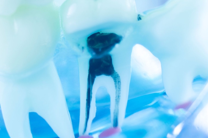
Conventional root canal therapy resolves endodontic infection by sacrificing the organ that suffered it. Once the pulp has been removed and replaced with gutta-percha and sealer, the tooth loses its ability to respond to stimuli, to produce reactionary dentin, to signal a carious or traumatic insult through pain and to mount a local immune defense against bacterial contamination. In immature permanent teeth the price is even higher, because the arrest of root development leaves thin walls and an open apex, with a fracture risk that no obturation material can compensate for. Regenerative endodontics arises precisely from this dissatisfaction, with the goal of restoring to the canal a living tissue that is vascularized, innervated and capable of dentinogenesis.
The clinical protocols most widely used today are based on revascularization through evoked bleeding. After disinfection of the root canal system, bleeding is induced from the periapical region, the resulting clot fills the canal and serves as a provisional scaffold for the stem cells of the apical papilla that migrate into it. Radiographic outcomes are often encouraging, with thickening of the canal walls and root lengthening, but histological analysis of extracted teeth tells a different story. The tissue that forms resembles cementum, bone and fibrous connective tissue far more than a true pulp with an organized odontoblastic layer. The clot is an unpredictable scaffold in terms of volume, composition and persistence, it does not retain growth factors in a controlled manner and it offers cells no mechanical cue consistent with the pulp phenotype. It is on this lack of control that biomaterials research has focused, and silk proteins occupy a particular position within it.
A microenvironment that imposes precise constraints
Before evaluating any scaffold, it is worth recalling how hostile the environment in which it must operate really is. The root canal is a low-compliance space, bounded laterally by mineralized dentin and accessible to the vasculature only through an apical foramen that in mature teeth measures fractions of a millimetre. Blood perfusion therefore arrives from a single direction, the oxygen gradient is steep and the core of any bulky construct tends toward hypoxia. Added to this are the residual biofilm that no irrigation protocol fully eliminates, the sodium hypochlorite residues that can oxidize surfaces and reduce cell adhesion, and EDTA conditioning, which besides removing the smear layer releases TGF-β1, BMPs and other signals sequestered in the dentin matrix during primary dentinogenesis.
A pulp scaffold must therefore be injectable, or at least adaptable to a thin and irregular geometry. It must have a stiffness compatible with that of loose connective tissue, which lies in the range of a few kilopascals, it must allow vascular invasion from the apex toward the pulp chamber and it must degrade with kinetics that leave room for the newly formed matrix without collapsing before that matrix can support itself. It must also tolerate the interface with conditioned dentin and, ideally, exploit the factors that dentin releases rather than masking them.
Fibroin as a tunable matrix
Regenerated fibroin lends itself to this set of constraints for reasons that anyone working with silk proteins knows well, but that take on specific weight in the endodontic context. The ability to modulate stiffness by acting on solution concentration, β-sheet content and the method used to induce the conformational transition makes it possible to obtain matrices ranging from soft gels to porous sponges while keeping the same underlying chemistry. Proteolytic degradation, slow compared with collagen and fibrin and adjustable through the degree of crystallinity and the chain molecular weight set during degumming, allows the persistence of the scaffold to be calibrated to the timescale of dentinogenesis, which is measured in weeks and months rather than days.
The first systematic work on pulp cells cultured on silk scaffolds came from Pamela Yelick's group in collaboration with David Kaplan's laboratory at Tufts University. As early as 2008, Xu and colleagues had used porous fibroin sponges seeded with porcine tooth bud cells, observing after in vivo implantation the formation of mineralized dentin-like and enamel-like tissues whose organization was influenced by pore size. In 2011 Zhang and colleagues compared the behavior of human dental pulp progenitors on scaffolds produced from aqueous solutions and from hexafluoroisopropanol solutions, two processing routes that yield different porous architectures, crystallinity and degradation rates. The comparison showed that the fabrication route is not a neutral technical detail, since it affects proliferation, cell distribution and the deposition of mineralized matrix. These studies yield an indication that still holds today, namely that in the pulp the choice of fibroin processing should be driven by the phenotype one aims to obtain and not by laboratory habit.
Injectable hydrogels and in situ gelation
Preformed porous sponges are useful for in vitro studies and ectopic implantation, but they can hardly adapt to a real root canal. For endodontic application the most coherent form is an injectable hydrogel that gels after placement. Fibroin offers several routes to achieve this. Sonication accelerates β-sheet assembly and allows gelation time to be tuned within a window of minutes, long enough to mix in cells or factors and inject the solution before it sets. Enzymatic crosslinking mediated by horseradish peroxidase and hydrogen peroxide, which exploits the tyrosine residues naturally abundant in the heavy chain, produces instead gels that are initially amorphous, elastic and very soft, and that over time spontaneously evolve toward higher crystallinity and therefore progressive stiffening. This behavior, which in other contexts may be a limitation, lends itself to an interesting reading in the pulp, because a matrix that starts soft favors migration and angiogenesis and then consolidates as the proliferative phase gives way to differentiation.
Shear-thinning formulations, obtained for example by blending partially assembled fibroin or combining it with other polymers, facilitate extrusion through small-gauge endodontic needles and the recovery of viscosity once deposited in the canal. Fibroin itself does not contain integrin-specific adhesion sequences in amounts comparable to collagen, and for this reason many groups turn to functionalization with RGD peptides or to blending with gelatin, collagen or hyaluronic acid to improve the adhesion and spreading of dental pulp stem cells and stem cells of the apical papilla within the gel.
Signal delivery and dentinogenesis
The real strength of fibroin in regenerative endodontics is its ability to stabilize and release bioactive molecules under physiological conditions. The repetitive hydrophobic domains and the β-sheet structure create microenvironments in which sensitive proteins such as VEGF, bFGF, PDGF and BMPs retain their conformation longer than in solution or in conventional hydrophilic matrices, and all-aqueous processing at room temperature avoids the organic solvents and temperatures that would denature the factors during loading. Release can be modulated by embedding the factors directly in the gel, by adsorbing them onto fibroin micro- and nanoparticles of different crystallinity dispersed in the matrix, or by combining the two strategies to obtain differentiated profiles.
This modularity matters because pulp regeneration requires a temporal sequence of signals. In the early phases a strong angiogenic and chemotactic stimulus is needed to recruit cells from the apex and ensure perfusion of the construct, while in later phases odontogenic signals prevail, driving the expression of DSPP, DMP-1 and the other markers of the odontoblastic lineage. Dual-release fibroin systems, with VEGF available rapidly and an osteo-odontogenic factor retained longer in highly crystalline particles, reproduce this sequence better than a blood clot or a matrix that releases its entire payload within the first hours. Fibroin is also well suited to carrying proteins extracted from the dentin matrix itself, an approach that exploits signals already naturally present in the tooth and reduces reliance on costly recombinant factors.
A less discussed aspect concerns the role of dentin conditioning. The TGF-β1 released by EDTA diffuses into the canal and can be intercepted by the scaffold. A fibroin hydrogel placed in contact with conditioned dentin tends to retain part of these factors in the layer closest to the wall, and it is precisely in that region that the polarization of odontoblast-like cells is expected. The gradient established between a periphery rich in odontogenic signals and a core dominated by any VEGF loaded into the gel mirrors, at least in principle, the organization of the native pulp with its peripheral odontoblastic layer and central vascular core.
The contribution of sericin
For a long time sericin was regarded as a by-product to be carefully removed, suspected of being responsible for the immune reactions attributed to silk. More recent studies have scaled back this interpretation, showing that the inflammatory responses observed derived mainly from the association of sericin and fibroin in the native fiber rather than from purified sericin as such. In the pulp context the properties of sericin take on specific interest, because the pulp to be regenerated arises in an environment that has been inflamed, often chronically, and that retains high oxidative stress even after disinfection.
Sericin has antioxidant activity linked to its high serine content and to residues bearing hydroxyl groups, it modulates the production of pro-inflammatory cytokines by macrophages and it supports the adhesion and proliferation of several types of mesenchymal cells. In vitro, adding sericin to fibroin scaffolds or preparing fibroin-sericin composite hydrogels has been shown to improve the viability of dental-derived stem cells and to attenuate the cytotoxic effects of oxidizing agents. Sericin is also far more hydrophilic than fibroin and, within a composite matrix, it accelerates hydration and nutrient diffusion in the first hours after implantation, exactly when the construct depends entirely on diffusion from the apex.
Its rapid degradation relative to fibroin is a limitation if it is used alone as a structural scaffold, but it becomes an advantage if it is regarded as a transient component that exerts a protective action in the early phase and then gives way to the fibroin matrix and the newly formed tissue. Variability related to the extraction method, which can yield fractions of very different molecular weight, remains a parameter to be controlled carefully, since it affects both biological activity and gel stability.
Controlling the bacterial load
Disinfection is the prerequisite of any regenerative procedure, and it also represents one of its paradoxes. Triple antibiotic paste and the calcium hydroxide preparations used as intracanal medicaments are effective against biofilm but, at traditional concentrations, they are toxic to the stem cells of the apical papilla that are supposed to repopulate the canal. Fibroin matrices offer a way to reduce this contradiction, because they allow antibiotics or antimicrobial agents to be released over a prolonged period at lower local concentrations, maintaining a bacteriostatic effect on the dentin surface without the cytotoxic peaks of conventional medicaments.
Fibroin films and electrospun nanofibers loaded with antibiotics have been studied, as have composites with silver nanoparticles or with naturally derived compounds with antimicrobial activity. Here too fibroin protects the carried molecule and regulates its diffusion according to its own crystallinity. Some approaches combine within the same matrix an antimicrobial compartment intended for the initial phase and a regenerative compartment that becomes active later, exploiting gel layering or particles of different stability.
Calcium silicate composites and pulp capping
Alongside regeneration of the entire canal there is a more conservative field, that of direct pulp capping and pulpotomy, in which the aim is to preserve the remaining pulp and induce the formation of a reparative dentin bridge. The clinical reference is calcium silicate cements, MTA and derived materials, which provide an excellent seal and release calcium hydroxide that promotes mineralization, but which have long setting times, handling difficulties and a very high initial pH that causes a superficial layer of necrosis.
Combining fibroin with calcium silicates, hydroxyapatite or bioactive glasses aims to obtain materials that retain the bioactivity of the mineral component while improving handling and reducing initial aggressiveness at the pulp interface. Fibroin acts as an organic phase that modulates ion release, dampens the alkaline peak and provides a substrate for pulp cell adhesion, while the inorganic component guides mineral nucleation. In animal model studies, dentin bridges have been observed with continuity and thickness comparable to or greater than those obtained with reference materials, together with more limited underlying pulp inflammation. Thanks to its ability to bind calcium ions through the acidic residues of the chain and to orient hydroxyapatite deposition, fibroin can take an active part in the mineralization process rather than merely acting as a carrier.
The dentin interface
The point that more than any other separates a good experimental result from a functional pulp tissue is the interface between matrix and dentin wall. A true pulp is not simply a vascularized connective tissue filling the canal, but a tissue in which odontoblasts align along the dentin, extend their processes into the tubules and deposit new predentin with a defined polarity. When the scaffold detaches from the wall, even by a few micrometres, this organization fails to form and the outcome drifts toward the disordered mineralized tissue observed in clot-based revascularization.
Fibroin has good affinity for conditioned dentin surfaces, thanks to interactions with the collagen exposed by EDTA and with residual mineral, and hydrogels that gel in situ fill wall irregularities better than a preformed scaffold. The contraction that accompanies increasing crystallinity over time is, however, a factor that must be measured and compensated, because it can open precisely the gap one sought to avoid. Formulations that maintain an elastic amorphous phase for longer, such as enzymatically crosslinked ones, or that include a composite component capable of bonding chemically to dentin, currently appear best suited to preserving the continuity of the interface during the weeks in which peripheral cells polarize and begin depositing matrix. It is on this detail, more than on the composition of the construct's core, that the difference is decided between a canal simply filled with living tissue and a pulp that once again behaves as one.
