
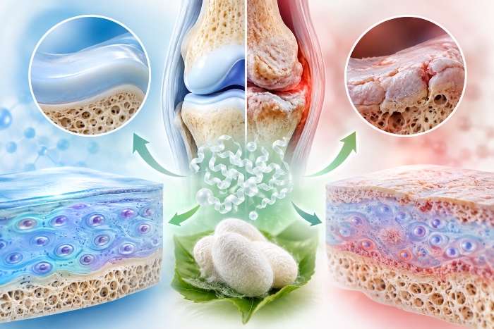
Osteoarthritis is not, as was long believed, the simple mechanical wear of a tissue destined to consume itself with age. It is a disease of the entire joint — cartilage, subchondral bone, synovial membrane, ligaments and menisci all take part in a dynamic, active process — whose common endpoint nonetheless remains the loss of hyaline cartilage. It affects more than two hundred and fifty million people worldwide, with the knee accounting for the heaviest share of the disability burden, and its prevalence continues to rise, driven by an ageing population and increasing obesity. Faced with a phenomenon of this scale, pharmacology still relies almost exclusively on symptomatic tools. Hence the growing interest in molecules capable not of masking pain but of intervening in the biology of the tissue itself: chondroprotective agents in the proper sense. It is in this space that sericin, a waste protein of the silk industry, is proving a surprisingly versatile candidate.
The architecture of a tissue that cannot repair itself
To understand why articular cartilage represents such a stubborn therapeutic challenge, one must begin with its structure. It is hyaline cartilage, a tissue made up of a single cell population, the chondrocytes, embedded in a dense and highly organised extracellular matrix. This matrix is the true functional tissue: its framework is provided by type II collagen (COL2A1), which supplies tensile strength, while the proteoglycans — with aggrecan as the protagonist and its long chains of glycosaminoglycans (GAGs) — retain water and give the tissue its capacity to resist compression. The chondrocytes, though they make up a minimal fraction of the volume, are the custodians of homeostasis: they continuously synthesise new matrix and, in equilibrium, regulate its degradation through dedicated enzymes.
The structural problem is twofold. Articular cartilage lacks blood vessels and innervation, and chondrocytes have reduced mitotic activity and live in a nutrient-poor microenvironment. The consequence is an almost non-existent regenerative capacity: a focal lesion does not heal spontaneously and tends, on the contrary, to progress, often evolving into post-traumatic osteoarthritis. Every therapeutic strategy must reckon with this underlying avascularity, which limits both the access of drugs and the possibility of recruiting reparative cells from outside.
The catabolic imbalance of osteoarthritis
In the healthy joint the chondrocyte maintains a fine balance between the anabolism and catabolism of the matrix. Osteoarthritis breaks this balance. The chondrocytes alter their behaviour, reducing matrix synthesis and increasing its breakdown: they begin to secrete pro-inflammatory cytokines — interleukin-1 beta (IL-1β), tumour necrosis factor alpha (TNF-α), interleukin-6 — which in turn induce the expression of degradative enzymes. Among these, the matrix metalloproteinases, in particular MMP-1 (collagenase-1) and MMP-13 (collagenase-3), efficiently attack type II collagen, while the aggrecanases of the ADAMTS family dismantle aggrecan.
The result is a self-sustaining cycle. The early alteration of the structure and composition of the GAGs within the proteoglycan is one of the initiating events of the process, followed by the degradation of type II collagen — the histopathological hallmark of the disease — and by the progressive reduction of the very number of chondrocytes. It is worth stressing the inverse relationship linking these markers: where IL-1β, TNF-α and MMP-1 rise, the expression of COL2A1 falls in a statistically significant way. Recovering type II collagen and switching off the inflammatory signature are therefore not two distinct objectives, but two faces of the same rebalancing.
What chondroprotection really means
The term "chondroprotective" is used with considerable looseness, often applied to supplements whose efficacy on the tissue is, at best, controversial. In the strict sense, a chondroprotective agent should do something more than relieve symptoms: it should preserve or reconstitute the cartilaginous matrix, contain the catabolic activity of the chondrocytes and, ideally, favour a stable cellular phenotype. This is the territory of the so-called DMOADs, the disease-modifying osteoarthritis drugs, a category that to this day remains largely a promise: no molecule has demonstrated incontrovertibly that it can halt cartilage loss in humans. Ordinary clinical management relies on analgesics, non-steroidal anti-inflammatory drugs, and intra-articular injections of corticosteroids or hyaluronic acid, while recourse to glucosamine and chondroitin remains a matter of debate. It is precisely this gap that makes any compound acting on the molecular mechanisms of the disease, rather than on its perceived effects, so interesting.
Sericin: from cocoon glue to bioactive protein
Sericin is a protein produced by the silkworm Bombyx mori. Within the cocoon it performs an apparently humble structural function: it is the glue-like substance that holds together the two fibroin filaments, forming the silk thread. In traditional silk processing it is removed and discarded, with a far from negligible environmental impact. But its amino acid composition tells a richer story. Sericin is dominated by polar amino acids — serine, threonine and aspartic acid — which endow it with marked adhesive properties and the capacity to form gels. These same chemical features have a biological correlate: the protein induces collagen production both in vitro and in vivo, shows a documented anti-inflammatory action with the suppression of mediators such as COX-2, iNOS and several interleukins, and possesses low immunogenicity, good biocompatibility and biodegradability. It is unsurprising, then, that in recent years it has migrated from the laboratories of dermatology and wound care towards experimental orthopaedics and cartilage tissue engineering.
What had long been missing was not evidence of a favourable effect of sericin on chondrogenic proliferation and viability — that already existed — but an understanding of the molecular mechanisms through which this effect is realised. It is on this ground that the most recent research has taken the decisive step.
The molecular pathways: glycolysis and TGF-β/Smad signaling
The most illuminating study in this direction is the work published in Scientific Reports in 2024 by the group of Fongsodsri and Ampawong, carried out on three-dimensional chondrogenic models of murine ATDC5 cells, both in pellet culture and on gelatin scaffolds. The authors first defined a non-cytotoxic concentration window, identifying doses of 1, 25 and 50 micrograms per millilitre as a low, medium and high level respectively, with cell viability maintained above eighty per cent up to 50 µg/ml. This methodological detail is already relevant: the beneficial effect we will see emerges most clearly at the highest concentration of the safe window.
The first result is metabolic in nature, and in part unexpected. The proteomic analysis identified more than a hundred upregulated proteins, and at the top of the list by variation in expression stand two glycolytic enzymes: phosphoglycerate mutase 1 (Pgam1), with an increase of roughly eighteen-fold, and triosephosphate isomerase (TPI), with an increase of roughly fourteen-fold. Glycolysis is the central metabolic pathway of the chondrocyte, a cell that lives in a hypoxic environment and depends on the anaerobic metabolism of glucose to produce energy. Sustaining glycolysis means sustaining proliferation, which is an energetically demanding process. It is telling, by contrast, that in human osteoarthritic chondrocytes it is precisely the glycolytic enzymes that are typically reduced: sericin appears to act in the direction opposite to the diseased phenotype, reigniting the cell's energetic machinery.
The second result concerns differentiation and its regulation, and it is here that the term "modulator" acquires its full meaning. Sericin does not merely push the cell towards maturation: it directs its trajectory. At the level of gene expression it produced a marked increase in Smad2 and Smad3, the transducers of the TGF-β pathway, and a simultaneous reduction in Smad1, BMP2, BMP4 and RUNX2, which belong to the BMP signalling branch. The distinction is anything but academic. The TGF-β/Smad2-3 pathway governs the homeostasis and development of cartilage and stabilises the SOX-9 protein, the master transcription factor of the chondrocyte, which in turn activates COL2A1 and the other genes of the cartilaginous matrix. The BMP branch, with RUNX2 downstream, drives instead towards hypertrophy and endochondral ossification — that is, towards the transformation of the chondrocyte into a cell that prefigures bone formation, an undesirable fate in articular cartilage. By suppressing RUNX2 through Smad2/3, the TGF-β pathway reduces matrix degradation; sericin, by favouring this branch at the expense of the other, steers the cell towards a stable and productive chondrocytic phenotype rather than towards terminal maturation.
This reading is consistent with the body of data. Sericin significantly increased the gene expression of SOX-9 and COL2A1, raised the production of glycosaminoglycans steadily from day seven to day twenty-eight — and it is worth noting that this occurred even in the absence of chondrogenic supplements, a sign that the protein can itself act as an inductive factor — and strengthened the organisation of the cytoskeleton, with an increase in F-actin and beta-tubulin, elements whose disorganisation is a feature of osteoarthritic chondrocytes. One detail refines the picture further: at the high concentration, sericin reduced over time the activity of alkaline phosphatase, a marker of chondrocyte hypertrophy. In other words, it not only favours collagen synthesis but appears to restrain the drift towards calcification. There remains, in fairness, a not entirely linear finding: aggrecan did not follow the same trend as collagen, showing instead a reduction in gene expression in one of the experimental conditions. This is an inconsistency that the authors themselves flag as deserving further study, and one that recalls how uncoupled the regulation of the individual matrix components can be.
The anti-inflammatory and anti-catabolic action
Chondroprotection is not exhausted in construction: it also requires the capacity to contain demolition. On this front the same study set up a model of early inflammation, stimulating the chondrocytes on scaffolds with IL-1β and TNF-α and then comparing sericin with two clinically significant references, dexamethasone and glucosamine sulfate. At the protein level the results were clear-cut: all concentrations of sericin significantly reduced the expression of IL-1β, TNF-α and MMP-1, while raising that of COL2A1 in parallel. More interesting still is the direct comparison: at the medium and high doses sericin lowered IL-1β more than glucosamine sulfate, and at the high dose it reduced TNF-α more effectively than glucosamine, approaching the profile of dexamethasone on several markers. The corticosteroid, as expected, reduced cytokines and collagenases, but sericin offered an advantage that dexamethasone does not possess and that glucosamine did not replicate: the ability to actively raise the synthesis of type II collagen, not merely to slow its loss.
It is necessary, however, to convey the limits of these data as well, because the scientific seriousness of the information depends on not omitting them. The robust anti-inflammatory effect was observed at the level of protein expression, whereas at the level of gene expression the differences in MMP-13 and COL2A1 did not reach significance relative to the untreated control. The discrepancy between protein and messenger suggests post-transcriptional regulatory mechanisms that remain to be clarified and cautions against overinterpreting the signal. These are, in any case, results obtained in vitro on three-dimensional models: a solid bridge towards the therapeutic hypothesis, not yet a demonstration of clinical efficacy.
Sericin as a scaffold: injectable hydrogels
Alongside its biochemical action on the cell, sericin possesses a second chondroprotective dimension, structural in nature, which makes it particularly suited to filling cartilaginous defects. Its propensity to form gels has been exploited to build biomaterials that mimic the extracellular matrix. The reference work is that of Qi and colleagues, published in Biomaterials in 2018, in which sericin was functionalised into sericin methacryloyl (SerMA) — by introducing methacryloyl groups onto the reactive amine and hydroxyl groups of the protein — so as to render it photo-crosslinkable. Injected in liquid form and then exposed to ultraviolet light, SerMA crosslinks in situ, forming a pure hydrogel through a minimally invasive procedure well suited to the joint environment.
The properties of this material are notable from an engineering standpoint. The hydrogel is adhesive to chondrocytes and promotes their proliferation even under nutrient-deprived conditions, a crucial aspect given the impoverished environment of cartilage. Its mechanical characteristics and its degradation rate can be tuned by varying the degree of methacryloyl substitution — with degradation rates at forty-five days measured, across the different formulations, at around seventy-three, forty-seven and thirty-seven per cent — allowing the scaffold to be adapted to the specific requirements of the repair. The material also displays an intrinsic photoluminescence that permits real-time monitoring of its status. But the most eloquent finding is the in vivo one: chondrocyte-laden SerMA hydrogels formed, eight weeks after implantation, artificial cartilage that molecularly resembled native cartilage. This line of research has continued towards composite materials, for example by combining sericin with graphene oxide to obtain a stiffness sufficient even for bone repair, confirming the versatility of the platform. On the specifically cartilaginous front, earlier work had already shown that scaffolds of collagen and sericin, enriched with hyaluronic acid and chondroitin sulfate, improve regeneration by favouring the deposition of the typical matrix components.
Why "modulator" is the right word
Setting these findings side by side, a coherent figure emerges, and one understands why speaking of sericin as a chondroprotective modulator is more precise than describing it as a mere regenerative or anti-inflammatory agent. A single-target drug acts on one node of the network; sericin intervenes on several axes simultaneously. On the metabolic plane it sustains the chondrocyte's glycolysis, supplying energy to proliferation. On the phenotypic plane it directs differentiation through the balance between the TGF-β/Smad2-3 pathway and the BMP/RUNX2 pathway, stabilising SOX-9 and the production of type II collagen while at the same time restraining the hypertrophic drift. On the anti-catabolic plane it reduces the inflammatory cytokines and the collagenases that dismantle the matrix. And on the structural plane, as a biomaterial, it offers a biomimetic, adhesive and biodegradable scaffold on which the cells rebuild tissue. It is the convergence of these actions — not any one of them taken in isolation — that defines an authentic chondroprotective profile, one that confronts osteoarthritis in its nature as a systemic imbalance of the joint rather than as a single symptom.
