
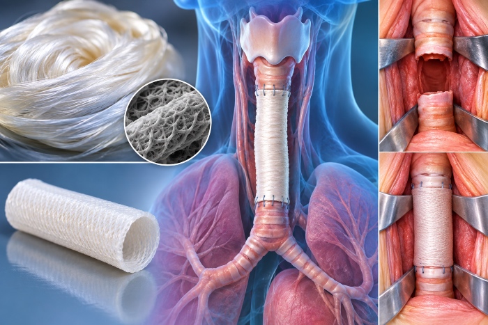
The trachea is a composite tube that must resist collapse under inspiratory negative pressure while at the same time retaining the longitudinal flexibility needed to accommodate neck movements and swallowing. Resection with end-to-end anastomosis is feasible up to roughly half the length of the organ in adults, beyond which the tension on the suture makes the outcome unreliable. For long defects the alternatives are limited, with allografts that require immunosuppression or complex revascularization procedures, and synthetic prostheses burdened by migration, granulation and obstruction.
An effective substitute must therefore fulfil three tasks simultaneously: supporting the wall with rings or structures analogous to the cartilaginous component, offering a lumen on which a functional ciliated epithelium can form, and integrating with an adequate blood supply to avoid ischemic necrosis of the mucosa.
It should be remembered that the diseases leading to extensive tracheal lesions are heterogeneous, ranging from post-intubation and post-tracheostomy stenoses to primary tumors such as adenoid cystic carcinoma, and extending to compressions and congenital malformations. This variety makes it difficult to establish a single product specification, and explains why much of the literature focuses on experimental segmental defects rather than circumferential ones, which are easier to reconstruct because they preserve a portion of native wall that guides epithelial regeneration.
Tubular configurations and fabrication processes
Fibroin lends itself to several strategies for building the tube. Knitted or braided structures provide radial strength and flexibility, while sponges obtained by gel spinning or freeze-drying confer interconnected porosity that is useful for cell colonization. Hybrid solutions, in which a textile weave is embedded in a porous matrix or combined with 3D-printed circumferential reinforcements, reproduce the ring-like anatomical design more faithfully. Extrusion printing of fibroin-based inks also makes it possible to modulate wall thickness and the distribution of reinforcements on patient-specific geometries derived from computed tomography.
The parameter that most often determines the outcome is the ratio between radial stiffness and compliance. A tube that is too stiff generates stress concentrations at the anastomoses, while one that is too compliant collapses during forced inspiration. Circumferential compression and bending tests should therefore be carried out under hydrated conditions and at physiological temperature.
Resistance to collapse depends non-linearly on geometry. A thin-walled tube with external spiral reinforcements or widely spaced rings can offer good radial resistance while maintaining flexibility, and three-dimensional printing makes it possible to optimize the pitch and thickness of the reinforcements. Compression tests between parallel plates and negative-pressure bench tests simulating the respiratory cycle provide more representative data than a simple measurement of the modulus of the bulk material, because it is the combination of geometry and material that determines the behavior of the implant.
Cell colonization and regeneration
Luminal re-epithelialization is the critical function, because ciliogenesis conditions mucociliary clearance and therefore the risk of recurrent infections. The approaches described involve seeding epithelial cells or mesenchymal stem cells on the luminal side and chondrocytes or chondrogenic progenitors in the wall component, or alternatively choosing acellular matrices that rely on endogenous recruitment. The latter route reduces regulatory complexity but exposes the patient to the risk of late and discontinuous epithelial coverage, with cicatricial stenoses.
In rabbit and swine models, good tissue incorporation is generally observed, but the lumen remains the weak point, with cases of narrowing due to granulation tissue. The use of a lumen pre-conditioned in a bioreactor, or revascularization by wrapping in omentum or fascia, are strategies that the literature continues to compare.
Pore size in the wall is a further delicate parameter. Pores that favor cellular and vascular infiltration may, if open toward the lumen, allow secretions and bacteria to penetrate the wall, and for this reason a gradient configuration with a more compact luminal surface is used. A low-porosity luminal membrane, possibly functionalized with extracellular matrix components such as collagen IV and laminin, can favor the adhesion and migration of the respiratory epithelium without compromising sealing.
