
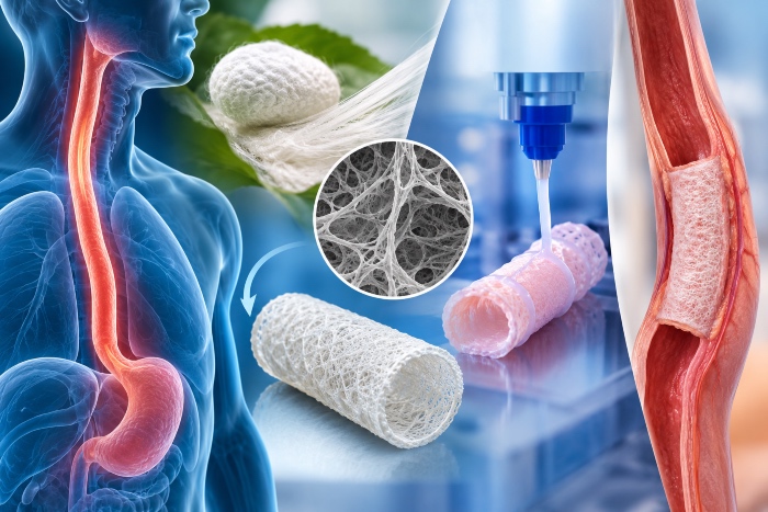
Among the organs of the digestive system, the esophagus is perhaps the one most easily underestimated. We picture it as a simple conduit, a muscular tube that carries the food bolus from the pharynx to the stomach, yet it is a surprisingly sophisticated structure. Within a few seconds it coordinates a peristaltic wave that runs through two different types of muscle, it protects itself from constant contact with food, saliva and at times gastric acid, and it does all this without the serosa that lines most other viscera. This very combination of functional complexity and anatomical fragility makes reconstruction of the esophageal wall one of the hardest challenges in surgery and, by extension, one of the most demanding test beds for tissue engineering.
Against this background, silk fibroin, the structural protein the Bombyx mori silkworm spins to build its cocoon, has emerged over the past decade as one of the most promising platforms for designing matrices able to guide esophageal regeneration from within. This is not a vague promise but a line of research that has produced solid experimental data in rodent and porcine models, together with a growing understanding of the molecular mechanisms that make tissue regrowth possible. It is worth retracing carefully, because it shows clearly both the potential and the still unresolved limits of this approach.
An organ that only looks simple
To understand why rebuilding the esophagus is so difficult, one has to start from its architecture. The esophageal wall is organised in concentric layers. On the inside lies the mucosa, lined by a stratified squamous epithelium that continuously renews itself from a population of basal progenitor cells. Next comes the submucosa, rich in vessels, glands and nerve plexuses, and then the muscularis propria, arranged in an inner circular layer and an outer longitudinal one. Here we meet a feature almost unique in the digestive tract, because in the upper segment the musculature is mainly striated, of the skeletal type, while further down towards the stomach it gradually gives way to smooth muscle. On the outside, instead of a serosa, there is only an adventitia of loose connective tissue, which makes sutures more vulnerable and anastomoses more prone to dehiscence.
On top of all this comes innervation. Esophageal peristalsis is not simply the result of muscle contraction but of a neural choreography involving the vagal system and the enteric nervous system, with the myenteric and submucosal plexuses synchronising contraction and relaxation along the entire conduit. A reconstructed tissue that fails to reinnervate properly may look intact under the microscope and still behave like a rigid, inert segment, unable to propel the bolus. This is exactly the kind of functional failure regenerative medicine must avoid.
The clinical problem and the limits of conventional surgery
The situations that call for replacing or repairing portions of the esophageal wall are varied and often dramatic. In newborns, esophageal atresia with a wide distance between the two ends, the so called long gap, prevents the ends from being joined directly. In adults, caustic ingestion, traumatic or iatrogenic perforation, refractory strictures and above all resections for cancer leave defects that cannot heal on their own.
The established surgical answer is to replace the missing segment with autologous tissue taken from elsewhere in the digestive tract, typically the stomach pulled up into the chest, or a segment of colon or jejunum. These are complex, life saving operations, but they come at a high price. Reconstruction with autologous gastrointestinal segments remains the gold standard, yet it is associated with complications such as dysphagia, donor site morbidity and, in some cases, patient death. Dysmotility adds to this, because tissue born to work as stomach or colon will never truly become esophagus, and it keeps its own secretory and contractile characteristics even in its new position.
Hence the need for ready to use materials, available without sacrificing another organ. Acellular matrices obtained from decellularised tissues, synthetic polymers or silk fibroin have been proposed precisely as off the shelf scaffolds to overcome the limits of autologous tissue. The principle is elegant. A cell free scaffold relies on the host's own progenitor populations, which must colonise the implant and generate new functional tissue. The matrix does not replace the organ, but offers the patient's cells a kind of temporary construction site in which to rebuild it, and then gradually disappears.
Why silk
Fibroin has a set of properties that make it particularly suited to this role. Its molecular structure alternates highly ordered hydrophobic domains, able to assemble into crystalline beta sheets, with more flexible amorphous regions. This architecture gives it remarkable mechanical strength combined with good elasticity, valuable qualities for a tissue that must withstand distension and compression thousands of times a day.
Then there is degradation. Fibroin is slowly broken down by the body's proteolytic enzymes and, by tuning the degree of crystallinity during processing, this process can be slowed or accelerated. In principle this makes it possible to synchronise the disappearance of the scaffold with the appearance of new tissue, a delicate balance that, as we will see, is still one of the critical points. Once separated from sericin through degumming, the protein also elicits a limited inflammatory response, and it is technologically highly versatile, since it can be turned into films, porous sponges, electrospun fibres, hydrogels and tubes.
This versatility is decisive for the esophagus, because it allows matrices that mimic its layered organisation. The most studied solution is the bilayer matrix, in which a compact fibroin film is annealed to a porous sponge of the same material. The film, facing the lumen, acts as a barrier against the leakage of saliva and food in the first weeks after implantation, when the new epithelium has not yet formed. The sponge, facing outwards, provides a network of interconnected pores into which muscle cells, fibroblasts, vessels and nerve fibres can penetrate and organise themselves.
The first laboratory tests
Before moving to animals, these matrices were tested in culture and compared with one of the biological materials most widely used in reconstructive surgery, porcine small intestinal submucosa, known as SIS. In these experiments fibroin matrices supported significantly greater attachment of smooth muscle cells of intestinal, colonic and esophageal origin than SIS. Both materials proved permissive for the contractile differentiation of these cells after stimulation with TGF beta 1, with increased expression of alpha smooth muscle actin and SM22 alpha, and both supported similar attachment and proliferation of esophageal epithelial cells.
Taken alone, these results say little in clinical terms, but they established an important premise. Fibroin is not merely tolerated by esophageal cells, it offers them an environment in which they can attach, multiply and acquire the contractile phenotype needed for function.
From rodent to pig, what happens in the body
The decisive step came with in vivo studies led mainly by Joshua Mauney's group, first at Boston Children's Hospital and later at the University of California, Irvine, in collaboration with David Kaplan's laboratory at Tufts University, one of the world's leading centres for silk as a biomaterial. The surgical technique used is onlay esophagoplasty, in which part of the esophageal circumference is removed and replaced with a matrix patch, leaving the rest of the conduit intact.
In the first rat study, esophagoplasty was performed with fibroin matrices in 40 animals, compared with 22 rats treated with SIS and 20 controls undergoing esophagotomy alone, for implantation periods of up to two months. Survival was 93 percent in the fibroin group and 91 percent in the SIS group, and all animals returned to solid food after three days on a liquid diet. Micro computed tomography at the end of the study showed no contrast leakage, fistulas, strictures or diverticula, and the reconstructed segments responded to contractile agents and electrical stimulation while relaxing under isoproterenol, a sign that the new tissue was not only present but working.
The most interesting finding, however, concerns the quality of regeneration. Fibroin matrices produced roughly a fourfold increase in skeletal muscle and a twofold increase in smooth muscle compared with SIS. In both groups a stratified squamous epithelium formed and new vessels and nerve endings appeared, but fibroin quadrupled the density of synaptic boutons in the implant region. Above all, SIS triggered chronic inflammatory reactions and severe fibrosis, which was not seen with silk. This is a highly relevant result, because fibrosis is precisely the process that leads to strictures, the most feared complication of any esophageal reconstruction.
The same group then set out to verify that the matrix would also work in an already damaged esophagus, a situation much closer to that of patients. In a caustic injury model, exposure to 40 percent sodium hydroxide caused corrosive esophagitis with a 91 percent reduction in luminal area. Repair with the bilayer matrix nonetheless achieved survival above 80 percent in both injured and healthy animals, with neuronal junctions and endothelium lined vessels forming at the graft site.
The leap to an animal closer in size to humans came with the pig, a model considered crucial in experimental surgery for its dimensions, anatomy and digestive physiology. Six adult pigs underwent thoracic onlay esophagoplasty with patches of about three by one and a half centimetres and were followed for three months. All survived without complications and resumed solid feeding, and esophagograms showed continuity of the organ with no leaks or strictures.
The hardest test, the circumferential defect
Repairing part of the wall while leaving the rest of the conduit intact is, however, very different from replacing an entire tubular segment. In the first case the surrounding healthy tissue supplies cells, vessels and nerves from every direction, as well as mechanical support. In the second, the matrix must hold the shape of the lumen on its own and receives cells only from its two ends.
To address this scenario, five Yucatan minipigs underwent reconstruction with two centimetre tubular bilayer grafts, combined with a temporary esophageal stent for two months followed by one month without it. The results were instructive precisely because they were not entirely positive. All animals survived and ate solid food, but in 60 percent of cases strictures developed at the graft site between the second and third month, requiring balloon dilation. At the same time, the matrices supported the formation of innervated, vascularised tubular neotissues with epithelial and muscular components capable of contraction and relaxation.
The most honest reading of these data is that fibroin works as a biological guide even in the tubular configuration, but is not yet enough to guarantee a conduit that remains stable over time. A 2024 review emphasised that the high stricture rate, together with insufficient innervation and poor peristalsis, makes the bilayer matrix not yet suitable for direct tubular esophagoplasty. Interestingly, the same kind of difficulty has emerged in urological applications, where in tubular ureteral reconstruction the authors concluded that in vivo degradation kinetics need optimising and stricture formation reducing. In other words, the problem is not specific to the esophagus but concerns the reconstruction of hollow conduits with acellular matrices in general.
The molecular mechanisms of re-epithelialisation
One of the most fascinating aspects of this line of research is the attempt to understand not only whether the matrix works, but why. Using mass spectrometry based quantitative proteomics and in silico pathway analysis, researchers identified the molecular cascades activated during formation of new epithelium in the rat after esophagoplasty with fibroin.
Experiments with pharmacological inhibitors gave a precise answer. Re-epithelialisation depends in part on survival signals that suppress caspase activity in epithelial progenitors, through activation of the hepatocyte growth factor receptor c-MET, the TrkA receptor, PI3K and Akt. Put simply, epithelial stem cells migrating across the matrix must receive a signal that keeps them from undergoing apoptosis, and that signal travels along a well defined molecular axis.
This finding opens concrete prospects for material design. If we know which pathways sustain regeneration, it becomes plausible to imagine functionalised silk matrices enriched with growth factors or molecules able to stimulate precisely those axes, exploiting fibroin's ability to incorporate and gradually release bioactive substances. It is a direction still to be explored experimentally, but it marks the shift from an empirical regenerative medicine to a rationally designed one.
Other architectures, electrospun fibres and basement membrane
Alongside bilayer matrices obtained by casting and freeze drying, other groups have explored electrospinning, a technique that produces networks of nanometric or micrometric fibres closely mimicking the fibrous organisation of the extracellular matrix. A review on electrospun fibroin had already discussed its potential applications in esophageal tissue engineering, highlighting its effect on the growth of smooth muscle cells, keratinocytes and fibroblasts.
A particularly refined approach concerns reconstruction of the basement membrane, the thin sheet on which the epithelium rests and which regulates its behaviour and differentiation. Some researchers prepared fibrous polylactide and fibroin scaffolds by electrospinning and coated them with basement membrane proteins extracted from porcine esophagus, including type IV collagen, laminin, entactin and proteoglycans. The idea is to offer epithelial cells not only physical support but also the biochemical language they recognise as their own, speeding the formation of a mature mucosal barrier. Composite materials that combine fibroin with degradable synthetic polymers are, moreover, a widespread strategy for tuning mechanical properties and resorption times more precisely.
