
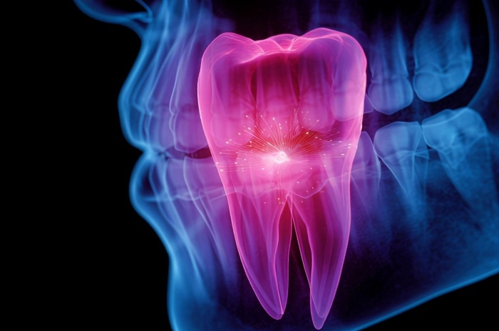
When the pulp dies and the tooth is still young
The dental pulp is a small organ, not a mere filling: a soft, richly vascularised and innervated connective tissue that nourishes the tooth, senses its stimuli, defends it against bacterial attack and, when needed, repairs damaged dentin thanks to the odontoblasts and to the stem cells that live in its perivascular niche. When deep caries or trauma lead to necrosis, conventional root canal therapy replaces the pulp with an inert material. In adults this is often sufficient. In children and adolescents, however, the problem is subtler: an immature permanent tooth still has an open apex and thin root walls, and the loss of the pulp interrupts the very process by which the root lengthens and thickens. The outcome is a tooth that survives but is fragile, exposed to fracture and to an uncertain future.
It is in this scenario that the idea arises of not merely filling but regenerating. The review on pulp regeneration strategies currently distinguishes three broad clinical approaches to pulp disease: root canal therapy, vital pulp therapy and regenerative endodontic therapy, the last of which aims to induce the formation of new functional tissue through autologous revascularisation or through pulp tissue engineering.
What regenerative endodontics really does, and where it stops
The term regenerative endodontic procedures was introduced by Murray in 2007 to stress the engineering essence of a treatment that had previously been called simply revascularisation. The American Association of Endodontists adopted this terminology in its guidelines, while the European Society of Endodontology speaks of revitalisation. The reference protocol, set out in the AAE clinical considerations, is aimed at teeth with a necrotic pulp and an immature apex: careful disinfection of the canal with 1.5 per cent sodium hypochlorite followed by a rinse with 17 per cent EDTA, induction of bleeding from the periapical tissues until the canal is filled with blood, and finally a seal with an MTA plug and a coronal restoration that prevents reinfection.
The blood clot is thus the scaffold, the framework on which stem cells from the periapical area are expected to migrate, adhere and proliferate. It is an elegant idea, but the clot is a material improvised rather than designed. The expert consensus published in 2022 points out that it is not always easy to obtain, that it lacks many of the properties of an ideal scaffold, such as ease of delivery, mechanical strength, controllable degradation and the possibility of incorporating growth factors, and that it contains numerous haematopoietic cells which, as they die, release toxic intracellular enzymes that can compromise stem cell survival. The clot may also contribute to tooth discolouration. Even the autologous alternatives, platelet-rich plasma and platelet-rich fibrin, have not convincingly been shown to improve outcomes compared with the clot alone.
There is also a conceptual knot. The AAE measures success on three levels, primary, secondary and tertiary, ranging from the healing of the infection through root development to the restoration of a vital response. But what grows back in the canal is not always authentic pulp: histological studies often suggest a repair tissue, closer to cementum or bone than to a pulp with organised odontoblasts, vessels and nerve fibres. Truly regenerating the dentin-pulp complex therefore requires a better scaffold than blood: one that is designed, disinfecting or at least compatible with disinfection, and capable of guiding the formation of vessels and of inducing odontogenic differentiation.
Silk as architecture: fibroin in pulp engineering
Bombyx mori silk is a two-component material. At the centre lie two filaments of fibroin, a hydrophobic protein rich in beta sheets that gives the fibre its strength and flexibility; around them is arranged sericin, a hydrophilic, adhesive protein that works as glue between the filaments. In industrial processing, sericin, which makes up roughly twenty to thirty per cent of the mass of silk, is removed by degumming and discarded as waste, in quantities estimated at around fifty thousand tonnes a year. In biomaterials laboratories, degumming, for instance with sodium carbonate, has long been an obligatory step to obtain a fibroin that is pure, crystalline, mechanically robust and of reduced immunogenicity.
Reviews devoted to pulp regeneration list clear advantages for fibroin: biocompatibility, biodegradability, the ability to carry drugs, high strength combined with flexibility, and the possibility of being processed as nanofibres, sponges or hydrogels. The disadvantages are equally clear, namely sensitivity to denaturation and some solubility constraints. A 2025 scoping review of scaffolds for the dentin-pulp complex mapped the literature and found that natural biomaterials and hydrogels appear in more than half of the included studies, with gelatin methacrylate, collagen, chitosan, hyaluronic acid and, indeed, silk fibroin among the protagonists.
The work that opened the way is that of Yang and colleagues in 2015. The group prepared porous fibroin scaffolds by freeze-drying, with or without basic fibroblast growth factor, seeded them with dental pulp stem cells and placed them in root fragments transplanted in an ectopic model. The cells survived and elongated within the scaffold for at least four weeks, and in the scaffold enriched with the growth factor a pulp-like tissue formed, well vascularised, with deposition of new matrix and the appearance of dentin-like tissue, made up of both transplanted and host cells. It was the proof of principle that a silk, suitably loaded, can support the reconstruction of pulp tissue.
From early prototypes to injectable hydrogels
The geometry of the root canal is an obstacle that is as much physical as biological: narrow, tortuous, irregular, hard to reach with a preformed scaffold. For this reason research has moved towards injectable hydrogels, which can fill any shape and solidify in situ. In this direction lies a photocrosslinkable composite hydrogel of methacrylated fibroin and methacrylated hyaluronic acid, in which human dental pulp stem cells showed good viability, proliferation and the capacity to differentiate along the odontogenic lineage.
A second line of work aims to solve the problem that limits regeneration more than any other, namely the poor oxygenation of an ischaemic and hypoxic canal. A 2025 study combined fibroin and sodium alginate in a hydrogel that releases, in a controlled way, apoptotic vesicles derived from dental pulp stem cells, known to promote the formation of microvessels. The results went beyond the in vitro setting: the construct accelerated vessel formation in a semi-orthotopic mouse model and, in an orthotopic beagle dog model, generated vascularised pulp tissue along the whole length of the canal, with aligned odontoblast-like cells and neuronal extensions. For regenerative endodontics, where vascularisation is the true bottleneck, this is one of the most encouraging findings in the silk literature.
Other groups have worked on the engineering of the scaffold itself. 3D-printed scaffolds of type I collagen and fibroin, with macropores of roughly 420 to 710 micrometres and porosity between 70 and 86 per cent, supported the proliferation and odontogenic differentiation of human pulp cells. Fibroin scaffolds enriched with graphene oxide enhanced the osteo-odontogenic differentiation of dental pulp stem cells. And a line of work of particular clinical interest concerns disinfection: methacrylated fibroin scaffolds loaded with antibiotics showed excellent cytocompatibility with stem cells from the apical papilla, comparable to that of pure scaffolds, positioning themselves as a delivery system able to control infection without harming the tissue that is meant to regenerate. This is a crucial point, because tissue growth stops where bacteria persist.
The other half of silk: sericin
If fibroin is the architecture, sericin may be the biology. For decades it was treated as a contaminant to be removed, because native sericin was associated with the immune reactions triggered by raw silk threads and its biosafety was long controversial. Today purified sericin is being reread in a very different light. The most recent reviews attribute to it antioxidant, anti-inflammatory and antibacterial activity and a capacity to promote cell proliferation, with low immunogenicity when properly extracted and purified. Antioxidant activity is regarded as the foundational property from which many others derive, and the antibacterial action has been linked to the presence of cysteine and its sulfhydryl groups. It must honestly be said that the strength of these properties depends heavily on purity and on the extraction method, and that sericin alone has poor mechanical properties.
For endodontics these attributes have an obvious resonance. A regenerating pulp lives in an environment of oxidative stress and residual inflammation, and its success depends on the ability to steer immune cells towards a reparative profile. Here sericin offers interesting clues, even though they come from other tissues. In scaffolds for periodontitis it reduced the expression of MMP-9 and MMP-3 and increased interleukin-10 in macrophages stimulated with lipopolysaccharide. In electrospun fibroin and sericin scaffolds at a seven to three ratio, used in muscle regeneration, it favoured the polarisation of macrophages towards the M2 phenotype and the formation of new vessels. Other studies report that sericin is able to recruit cells that promote regeneration, including endothelial cells. These are exactly the mechanisms that a regenerating pulp needs, even though none of these studies was carried out in the root canal.
On the pulp itself, in fact, there is at least one direct piece of evidence, and it is candid in showing both promise and limits. In a preliminary histological study on Wistar rat molars, sericin was used as a direct pulp capping material and compared with calcium hydroxide. At seven days calcium hydroxide produced less necrosis and less inflammatory infiltrate; at thirty days, however, the sericin-treated group showed a reduction in inflammation over time and a proliferation of new cells. The authors concluded that sericin improves the inflammatory response of the pulp but, applied in pure form, is not able to induce tertiary dentin formation. It is a result that does not delegitimise sericin: it places it rather as a modulator of the environment, more than an inducer of mineralisation, and therefore as a natural partner for fibroin and for odontogenic signals.
Purity, dosage and the question of immunogenicity
Here lies the question that runs through the whole field: is it better to have a degummed fibroin, pure and predictable, or a silk that retains a controlled fraction of sericin? Reviews of immune responses to silk biomaterials point out that fibroin lacks the antibacterial and antioxidant properties of sericin, but that both proteins, when purified, show low immunogenicity and good immunomodulatory properties. They also point out the practical limits: extraction and purification processes are complex and can leave residues and alter the properties of the material. Any protein material, after all, can in principle trigger an immune reaction, and the ratios between the two proteins must be demonstrated and not assumed.
The muscle data on the seven to three ratio is, in this sense, instructive: it suggests that there may be a compositional window in which sericin ceases to be a risk factor and becomes a tool for directing the immune response. Whether such a window exists for the pulp as well is an open question, and is probably the most promising research point of the entire subject.
Scaffold, signals and infection control
Putting the pieces together, the picture that emerges is that of a silk that solves different problems with different components. Fibroin provides a three-dimensional, porous body, injectable once functionalised, capable of housing cells and of releasing growth factors, vesicles or antibiotics in a programmed way. Sericin, around or within that structure, could provide a bioactive fraction with antioxidant and anti-inflammatory effects, a contribution to macrophage modulation and a possible aid in countering bacterial contamination. All this with a view to overcoming the blood clot as a random scaffold and arriving at a truly regenerated pulp, with vessels and nerves, and not merely a repair tissue.
Equally important is to state what we do not know. Almost all the evidence on fibroin in regenerative endodontics comes from in vitro studies and animal models, and, as far as emerges from the literature consulted, there are no human clinical trials of silk scaffolds for pulp regeneration. For sericin the picture is even subtler: the only direct evidence on the pulp is a preliminary study on a few animals, in which the pure material did not induce dentin, while the strongest arguments come by analogy from other tissues. The practical matter of standardisation also remains open: purity, molecular weight, batches, sterilisation, material stability and behaviour in the presence of hypochlorite, EDTA and intracanal medicaments.
