
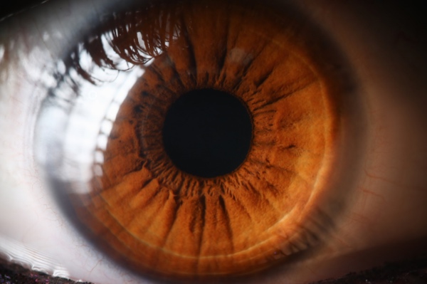
The cornea is the most transplanted tissue in the world and, thanks to the immune privilege of the anterior segment, the one with the best prognosis of any human graft. That very success makes the gap between those who could benefit and those who actually get access all the more jarring. The most frequently cited estimate, drawn from the global survey by Gain and colleagues in JAMA Ophthalmology, puts roughly 12.7 million people on the waiting list for keratoplasty, with a single cornea available for every seventy needed and more than half the world's population effectively without access to donor tissue. On top of this, about a third of the corneas procured each year are discarded as unsuitable, and donated tissue has a usable window of only a few weeks. It is the kind of problem no expansion of eye banking can solve on its own, and it pushes research toward a corneal substitute that can be manufactured, stored and distributed at global scale, independent of donation. In this landscape silk fibroin has emerged as one of the most studied materials, and the reasons are written entirely in the physics of how it is processed.
What an artificial cornea has to do
Before assessing a material it helps to fix the functional specification it must meet, because the cornea is an optical and a mechanical device at once. Its transparency is not a passive property but the result of the orthogonal organization of stromal collagen lamellae, spaced with sub-wavelength regularity so that scattering cancels out by interference. That same architecture provides biomechanical strength and curvature stability, which account for roughly forty of the eye's sixty diopters of refractive power. Transparency must also be actively maintained, because the stroma tends to swell and cloud if the endothelium does not pump out its excess water, and the whole tissue is avascular, therefore nourished by diffusion from the aqueous humor. A credible substitute must therefore be optically clear in the visible range, permeable to glucose and metabolites, mechanically compatible with suturing or intrastromal implantation, capable of supporting the adhesion and correct phenotyping of corneal cells, and not least immunologically silent. The tissue's stratification complicates the picture further, because epithelium, stroma and endothelium impose different and partly contradictory demands on the same biomaterial.
Why fibroin is a credible candidate
Fibroin's advantage in this context is that the properties that matter are controlled upstream, at the processing stage. The transparency of solvent-cast films can be tuned by choosing the starting solvent, from aqueous to formic acid to hexafluoroisopropanol, and by adjusting the annealing treatment with water vapor, methanol or steam, steps that govern conversion toward the β-sheet conformation. It is precisely this degree of secondary-structure crystallinity that simultaneously sets three parameters that are critical in the cornea and normally hard to decouple: optical clarity, mechanical stiffness and rate of proteolytic degradation. Raising the crystalline fraction yields films that are more stable and degradation-resistant but stiffer; lowering it gains ductility at the cost of persistence. Having a single processing variable that commands the whole triad is a rare advantage, and it explains why recent studies have mapped the process–property–function relationship of fibroin films specifically for corneal use, showing visible-range transmittance comparable to native tissue and elastic moduli tunable across a useful range. To this is added format versatility, from films to gels, sponges and nanoparticles through to 3D-printing bioinks, which lets the same material be cast into different geometries for different corneal layers.
The epithelium and limbal stem cells on a fibroin membrane
The first layer tackled successfully was the epithelium, and not by chance. The amniotic membrane has long been the substrate of choice for expanding and transplanting limbal epithelial stem cells in limbal deficiency, but it carries batch-to-batch biological variability, dependence on donation and a residual, if controlled, transmission risk. A fibroin film offers a defined, reproducible surface free of these constraints, on which limbal cells adhere, proliferate and stratify while retaining stem-phenotype markers. The ability to functionalize the surface with adhesion motifs or micro-structure it to orient growth turns the fibroin membrane into a programmable cell carrier rather than a merely inert support. On this front fibroin does not have to replace an entire stroma but a thin, transparent sheet that delivers cells, which is exactly the setting where the material performs best.
The stroma the real biomechanical proving ground
The stroma remains the hardest obstacle, because it makes up about ninety percent of corneal thickness and its transparency depends on a hierarchical lamellar organization that no biomaterial faithfully replicates today. Fibroin strategies diversify here: thin films stacked to reconstitute a stratification, porous sponges to house keratocytes, aligned fibers to mimic lamellar anisotropy, 3D-printed constructs to control fine-scale geometry. A particularly instructive line is that of blends, in which fibroin is combined with collagen to correct its best-known limitation: collagen is the native protein of the stromal matrix, hence maximally biomimetic, but mechanically weak and in need of crosslinking; fibroin supplies the intrinsic robustness and stability collagen lacks, and several works have shown how adding fibroin improves the mechanical properties of collagen-based membranes designed for corneal engineering. The challenge is not obtaining a transparent, resistant material, now within reach, but reproducing the stroma's biomechanical anisotropy and its long-term dimensional stability under physiological hydration.
The endothelium and the topographies that steer the pump phenotype
The endothelium is the clinically most consequential layer, because its inability to regenerate in humans makes Fuchs endothelial dystrophy the leading indication for transplantation in many parts of the world. Here fibroin works at a different scale, that of surface topography. Studies using fibroin from non-mulberry species as well, such as Antheraea assamensis, have shown that adding hexagonal micro-patterns or microgrooves to films modulates the behavior of corneal endothelial cells, guiding their mosaic arrangement and favoring the pump phenotype that keeps the stroma dehydrated. In parallel, characterization of Young's modulus and transmittance confirms that these microstructures do not compromise the substrate's optical and mechanical properties. A micro-structured fibroin film acting as a carrier for a cultured endothelial monolayer would open a route to the numerically heaviest indication, bypassing dependence on donor tissue precisely where the shortage is most acute.
From films to photocrosslinkable hydrogels and 3D printing
The most recent evolution shifts attention from the rigid film to formats that engage directly with the ocular surface. Photocrosslinkable fibroin hydrogels can be photobonded to the rabbit cornea as an alternative to the amniotic membrane in treating abrasions and ulcers, with a sutureless, minimally invasive approach that reduces surgical trauma and exploits fibroin's characteristic degradation tunability. On the reconstructive side, fibroin bioinks for 3D printing allow layer-by-layer control of construct geometry, with the prospect of building lamellar inlays or full-thickness constructs that trace the native architecture. It is the conceptual move from a material that mimics the cornea to a process that fabricates it to measure, and it is the direction in which fibroin's processability shows its most mature potential.
