
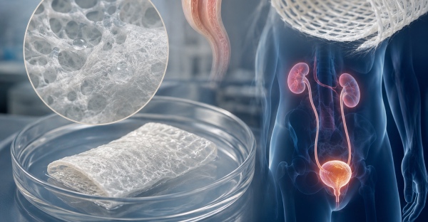
The lower and upper urinary tracts share a common underlying problem. When a segment of urethra, bladder or ureter is lost to recurrent strictures, trauma, oncological outcomes, congenital malformations or fibrotic processes, the residual tissue rarely provides enough material for a tension-free reconstruction. Classical reconstructive medicine has answered by turning to autologous tissues, from buccal mucosa in urethroplasty to detubularised intestinal segments in bladder augmentation. These solutions work, yet they carry significant morbidity with them. Enterocystoplasty in particular introduces into the urinary reservoir a mucus-secreting epithelium never designed to hold urine, with well-documented consequences ranging from excessive mucus production to metabolic disturbances, from recurrent lithiasis through to a long-term neoplastic risk.
This is where the interest in an engineered alternative arises. The goal is not simply to plug a defect but to provide a temporary matrix that mechanically supports the reconstructed region, withstands hydrostatic pressure and the dynamics of filling and voiding, and is meanwhile progressively colonised and replaced by functional autologous tissue. For this to happen the biomaterial must possess a property profile that few candidates manage to express simultaneously, and this is precisely the ground on which fibroin has proven its worth.
Fibroin as a structural platform
The fibroin extracted from the cocoon of Bombyx mori is a fibrous protein whose molecular architecture, dominated by crystalline beta-sheet regions embedded in amorphous domains, gives the material a strength-to-toughness ratio rarely matched by natural polymers. In urology this translates into a practical feature of enormous relevance, namely the possibility of producing thin, workable scaffolds that nonetheless hold sutures, resist intraoperative traction and maintain their integrity under cyclic load. A urethral or bladder graft must not only be biologically permissive, it must also be manipulable by the surgeon and able to retain stitches without tearing, and fibroin meets this requirement with margin to spare.
The second pillar is biocompatibility. Once the more immunogenic sericin components have been removed through degumming, fibroin evokes a contained and predictable inflammatory response, with a macrophage recruitment that tends to steer towards a pro-regenerative rather than a pro-fibrotic phenotype when the scaffold architecture is correctly designed. This aspect is crucial in the urinary tract, where an exuberant inflammatory response almost inevitably translates into fibrosis, cicatricial retraction and, in the case of the urethra, into stricture recurrence.
The third element is the controllability of degradation. Fibroin is degraded proteolytically, and the rate of the process can be modulated by acting on beta-crystallinity content, porosity, thickness and post-forming treatment strategies. This makes it possible to design a scaffold that persists long enough to accompany the regeneration of the wall, without remaining indefinitely as a foreign body exposed to urine, a condition that would favour encrustation and bacterial colonisation.
Scaffold architecture for the urinary environment
Designing a urological fibroin scaffold does not end with the choice of material, because it is the architecture that determines its biological fate. One of the most studied paradigms is the bilayer matrix, conceived to functionally reproduce the stratification of the urinary tract wall. The typical configuration features a dense, relatively compact luminal surface, obtained by surface fusion of the fibroin film, which acts as a barrier against urine extravasation and reduces the permeation of toxic solutes towards the surrounding tissues in the early phases of healing. Coupled to this is a porous, spongy outer layer, generated through lyophilisation or porogens, which offers the three-dimensional space needed for cell infiltration, matrix deposition and neovascularisation.
This duality resolves an otherwise irreducible design tension. A perfectly dense material would contain urine but would remain impermeable to cellular colonisation, whereas an entirely porous material would favour ingrowth yet would let urine seep into the perilesional tissues, triggering inflammation and fibrosis. The bilayer structure decouples the two functions by assigning them to distinct compartments of the same construct.
Alongside bilayer films, forming technologies have produced tubular structures for the replacement of urethral or ureteral segments. Gel spinning and electrospinning make it possible to fabricate conduits with a graded-porosity wall, while the cylindrical geometry can be produced on mandrels of defined calibre to match the desired luminal diameter. The ability to simultaneously control internal calibre, wall thickness and microporosity makes fibroin particularly versatile for the reconstruction of hollow structures, where luminal patency over time is as important as wall regeneration.
Urethral reconstruction and urethroplasty with fibroin matrices
Urethral stricture is perhaps the most demanding testing ground for a biomaterial, because recurrence is governed by fibrosis and every failure tends to worsen the tissue bed available for the next attempt. Fibroin scaffolds have been studied both in an acellular configuration, as an onlay matrix or as a tubular substitute, and in a cellularised version seeded with autologous urothelial and smooth muscle cells. Preclinical models, conducted predominantly in rodent and rabbit, have documented the regeneration of a stratified urothelium on the luminal surface and the appearance of organised smooth muscle bundles in the deeper layer, with a degree of fibrotic deposition generally lower than that of comparison biomaterials.
The clinically most relevant finding to emerge from this literature concerns luminal calibre at follow-up. Bilayer fibroin tends to preserve the lumen better than purely porous matrices, plausibly because the dense layer limits early cicatricial contraction and urine leakage during the most vulnerable phase of healing. In the perspective of substitution urethroplasty, where failure coincides with restenosis, this property represents a concrete advantage rather than a merely histological one.
The question of critical length remains open. As with all acellular scaffolds, there is a limit beyond which centripetal regeneration from the native margins fails to colonise the entire construct before fibrosis or luminal collapse set in. Overcoming this limit requires either precellularisation of the construct or strategies to accelerate neovascularisation, themes on which research is actively concentrated.
Bladder regeneration and augmentation
Engineered bladder augmentation is the application where fibroin has produced some of the most encouraging preclinical results. In cystoplasty models in rat and pig, bilayer fibroin matrices implanted as a patch replacing a defect in the bladder wall have shown regeneration of all three functional layers of the wall, from urothelium to muscle coat, accompanied by neovascularisation and a progressive innervation of the neotissue.
The parameter that makes these data particularly interesting is functional before it is morphological. Bladder capacity and compliance, meaning the reservoir's ability to accommodate increasing volumes of urine without pathological pressure rises, were in several studies maintained or recovered, a sign that the neotissue does not merely close the defect but participates in the mechanics of the organ. This is exactly what is missing from intestinal-segment solutions, which introduce a tissue structurally and metabolically foreign to the function of urinary containment.
The direct comparison between fibroin and acellular small intestinal submucosa, historically one of the reference materials in bladder regeneration, has on several occasions shown for fibroin a lower graft contraction and a more organised muscle deposition. The advantage appears to lie in the greater mechanical stability and the more predictable degradation kinetics of the silk protein, which offers the host tissue a more controlled temporal window for remodelling.
Ureteral reconstruction
The ureter poses a peculiar geometric and biomechanical problem, because it is a long, thin and peristaltic conduit whose function depends on luminal patency and on fine muscular coordination. The replacement of extended ureteral segments remains one of the unsolved problems of reconstructive urology, and tubular fibroin constructs have been explored as a possible answer. Fibroin conduits, often temporarily stented to ensure drainage in the initial phase, have shown in animal models urothelial regeneration and partial recovery of the wall structure.
The dominant criticality remains anastomotic stenosis and long-term durability. Ureteral peristalsis, the active component of urine transport, is difficult to reconstitute because it requires not only organised smooth muscle but also a coordinated innervation, an objective that no scaffold, whether of silk or of any other material, has so far achieved in a fully satisfactory way. Fibroin offers a mechanically adequate substrate, but clinical translation in this district is the least mature among those discussed.
Cellularisation, vascularisation and innervation
The distinction between acellular scaffold and cellularised construct runs through the whole of urinary tract engineering. The acellular approach is surgically simpler, avoids the complexities of cell culture and reduces costs and timelines, but it entrusts regeneration to the migration of native cells from the margins, with the dimensional limits already recalled. Precellularisation, which seeds the scaffold with autologous urothelial and smooth muscle cells before implantation, accelerates the formation of functional tissue and widens the length of treatable defect, at the price of a far more complex regulatory and manufacturing pathway.
Whatever the strategy, the common bottleneck is vascularisation. A construct thicker than a few hundred microns cannot rely on diffusion alone to nourish the cells within it, and without a capillary network establishing itself rapidly the centre of the scaffold undergoes necrosis, followed by fibrosis. Research addresses the problem by functionalising fibroin with angiogenic factors, VEGF in particular, exploiting the ability of the silk matrix to bind and release bioactive molecules in a controlled manner. Innervation, necessary for coordinated contractility above all in the bladder and ureter, remains the most arduous and least controllable goal, dependent on signals that the scaffold can favour but not dictate.
Degradation, encrustation and resistance to the urinary environment
Urine is anything but a neutral fluid from the material's point of view. Its ionic composition favours the precipitation of salts and the formation of encrustations on exposed surfaces, while the urinary tract is subject to bacterial colonisation and recurrent infections that alter the local pH and amplify the risk of lithiasis on the biomaterial. An ideal urological scaffold should therefore degrade on a timescale synchronised with urothelial regeneration, so that the surface exposed to urine is rapidly covered by a protective native epithelium before encrustation or infection take hold.
Fibroin lends itself well to this balancing act because its degradation rate is governable upstream. By increasing beta-sheet crystallinity, for example through water-vapour annealing, proteolysis is slowed and the persistence of the scaffold is prolonged, whereas lower crystallinity accelerates resorption. The design challenge lies in finding, for each district, the temporal window that allows complete urothelial coverage before the mechanical integrity of the matrix is compromised.
Antimicrobial functionalisation and drug release
One of the most valuable properties of fibroin in urology is its nature as a pharmacological reservoir. The matrix can incorporate and sustainedly release molecules of different nature, and this opens the way to scaffolds that do not confine themselves to providing structural support but exert an active therapeutic function. In the context of urethral strictures, where fibrosis is the engine of recurrence, incorporating antifibrotic agents such as mitomycin C within the construct represents a logical research direction, since it allows a local and prolonged release exactly where it is needed, avoiding systemic exposure.
On the infective front, antimicrobial functionalisation of the scaffold, whether through metallic nanoparticles or through antimicrobial peptides bound to fibroin, aims to counter bacterial colonisation during the phase in which the construct surface is still exposed and most vulnerable. The ability of fibroin to stabilise bioactive molecules without denaturing them, together with its gradual degradation, makes it a particularly suitable vehicle for these long-term releases, and sets these constructs apart from mere passive wall substitutes.
