
Pathophysiology of disc degeneration and biomaterial rationale
The intervertebral disc is a highly specialized fibrocartilaginous structure that fulfils primary mechanical functions: axial load dampening, compressive force distribution, and conferral of mobility to the spine across all planes of movement. Its architecture is tripartite — nucleus pulposus (NP), annulus fibrosus (AF), and cartilaginous endplates — and each of these compartments is governed by a biochemical and biomechanical microenvironment profoundly distinct from the others. Intervertebral disc degeneration (IDD) presents as a progressive and multifactorial process in which the loss of the NP extracellular matrix (ECM) — dominated by aggrecan, chondroitin sulphate, and hyaluronic acid — leads to a reduction in intradiscal osmotic pressure, vertebral height collapse, anomalous redistribution of loads onto the AF and facet joints, and ultimately segmental instability accompanied by chronic pain. At the cellular level, the phenotypic transition of notochordal cells toward a quiescent chondrocytic profile, progressive hypoxification due to the absence of direct vascularisation, and the accumulation of advanced glycation end-products (AGEs) alter ECM homeostasis by reducing the synthesis of proteoglycans and type II collagen in favour of type I collagen — a well-established marker of pathological fibrocartilaginisation.
Within this pathophysiological framework, tissue engineering strategies converge on the need for a scaffold material capable of replicating the NP microenvironment: high water content, non-linear viscoelasticity dependent on loading frequency, swelling capacity in response to osmotic gradients, biocompatibility with hypoxic cells at low cellular density, and the possibility of functionalisation with bioactive molecules or morphogenetic signals. Fibroin, by virtue of its intrinsic structural and rheological characteristics, lends itself exceptionally well to satisfying this complex set of requirements.
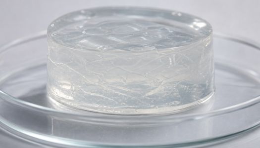
Structural and rheological properties of fibroin hydrogels relevant
Fibroin in aqueous solution — in its random coil or alpha-helical conformation — can be induced to form three-dimensional hydrophilic networks through several gelation mechanisms: thermally or ultrasonically induced β-sheet self-assembly, physical crosslinking mediated by methanol or ethanol, chemical crosslinking with bifunctional agents such as glutaraldehyde or genipin, and photopolymerisation of methacrylated derivatives (SilkMA). Each of these approaches generates a hydrogel with markedly different mechanical properties, porosity, and degradation kinetics, thereby enabling fine-tuned formulation tailored to the target compartment.
For NP applications, the reference rheological parameters include a compressive modulus in the 1–10 kPa range, viscoelastic behaviour with a loss tangent (tan δ) between 0.1 and 0.3 across physiologically relevant frequencies (0.01–10 Hz), and an equilibrium osmolar swelling capacity sufficient to maintain an intradiscal pressure of 0.1–0.3 MPa under static loading conditions. Pure fibroin hydrogels achieve storage moduli (G') typically ranging from 0.5 to 20 kPa depending on protein concentration (commonly 5–20% w/v), degree of β-crystallinity, and crosslink density, placing them in a range compatible with the healthy nucleus pulposus.
A particularly noteworthy rheological feature is the selective hydrophobicity of fibroin: crystalline domains rich in GAGAGS sequences form insoluble β-sheets that act as permanent physical junctions, while glycine-rich amorphous domains preserve chain mobility and hydraulic permeability. This duality confers an adaptive mechanical behaviour on the hydrogel — resistant to rapid compression, viscous under slow loading — that faithfully mimics the non-linear response of the native NP. Modulation of the ratio between crystalline and amorphous fractions, achieved by controlling treatment time and temperature or the concentration of denaturing solvents, constitutes a critical lever in the experimental design of optimised formulations for intradiscal use.
Functionalisation strategies and bioactive molecule release
One of the most significant advantages of fibroin hydrogels over other natural biomaterials (alginate, gelatin, collagen) is the abundance of reactive side-chain groups — carboxyls, amines, hydroxyls — that can be exploited for covalent conjugation of adhesive peptides, growth factors, glycosaminoglycans, or pharmacological ligands without significantly compromising the mechanical properties of the network. In the disc context, the molecules of greatest applicative interest are TGF-β1, GDF-5 (Growth Differentiation Factor 5), bFGF, and IGF-1, all of which possess documented chondroprotective activity on NP cells.
The incorporation of TGF-β1 into fibroin matrices has been achieved through simple physical entrapment within the polymer network (encapsulation), through biotinylation of fibroin and streptavidin-mediated immobilisation, and through covalent conjugation via PEG-based biodegradable linkers. The resulting release profiles depend critically on crosslink density and the presence of macroporous channels: high-crystallinity fibroin hydrogels exhibit a biphasic release with an initial burst of approximately 20–30% within the first 24 hours, followed by near zero-order kinetics sustained over weeks — a profile compatible with the chronobiology of the disc regenerative response. Another particularly promising line of functionalisation for disc applications involves the conjugation of hyaluronic acid (HA) to fibroin, achieved through click chemistry or carbodiimide coupling agents. HA contributes three synergistic effects: enhancement of osmotic swelling capacity, activation of the CD44 receptor expressed by NP cells (with anti-apoptotic downstream signalling mediated via the PI3K/Akt pathway), and negative modulation of inflammation through NF-κB suppression induced by high-molecular-weight HA fragments. Fibroin–HA composites reach equilibrium water contents exceeding 90% under isotonic conditions, approaching closely the values of the healthy NP (80–90%), and maintain adequate compressive moduli even under prolonged cyclic deformation.
Cellularisation and co-culture: biological integration of the hydrogel
An acellular hydrogel, however biomimetically sophisticated, encounters intrinsic limitations in a hypoxic, avascular, and nutrient-poor microenvironment such as that of the disc. The integration of mesenchymal stem cells (MSCs) derived from bone marrow, adipose tissue, or Wharton's jelly within the fibroin matrix prior to gelation represents the most extensively investigated strategy for conferring active regenerative capacity to the implant. MSCs encapsulated in fibroin hydrogels maintained under low oxygen tension (1–2% O?, reproducing the NP microenvironment) and stimulated with GDF-5 and TGF-β3 display a chondrocytic phenotype marked by upregulation of SOX9, ACAN, and COL2A1, with concurrent downregulation of COL1A1 — a differentiation trajectory toward the notochordal-like phenotype of the mature NP.
Long-term cell survival depends critically on the interconnected porosity of the hydrogel, which governs the diffusion of oxygen and glucose toward the core of the construct. Engineering porosity in fibroin hydrogels can be achieved through several techniques: controlled lyophilisation followed by rehydration, salt leaching, biofabrication via 3D extrusion-based bioprinting of fibroin bioinks or UV stereolithography of SilkMA derivatives, or the creation of interpenetrating network (IPN) structures with degradable porogen polymers. In optimised formulations, an interconnected pore density in the 50–200 µm range is sufficient to ensure cell viability above 80% for up to 21 days under static conditions, and up to 28 days with perfusion bioreactors. Co-culture of MSCs with primary NP cells within the same fibroin matrix — or in transwell systems that mimic native compartmentalisation — has demonstrated synergistic paracrine effects: NP cells secrete CXCL12 and CCL5 that enhance MSC recruitment toward the matrix, while MSCs release TSG-6 and PGE? that suppress the pro-inflammatory profile of degenerative NP cells, reducing the production of IL-1β, TNF-α, and MMP-3.
Preclinical models and in vivo biomechanical data
Preclinical validation of fibroin hydrogels for disc regeneration has developed primarily in rat-tail animal models (nucleotomy or needle puncture as degeneration-induction methods), rabbit, and sheep, with a growing number of studies in porcine ex vivo models that offer disc geometry and loading profiles closer to those of humans. Across all these models, the primary evaluation parameters include the disc height index (DHI), T2-weighted MRI signal as a proxy for NP water content, biochemical content of proteoglycans and type II collagen on histological analysis, and biomechanical assessment of disc compliance under axial compression and flexion.
In rat studies (21G needle puncture model at the caudal C5-C6 level), intradiscal injection of GDF-5-loaded fibroin–HA hydrogel showed, at 8 weeks post-treatment, a DHI of 78–85% relative to baseline versus 52–58% in the placebo group, and a normalised T2 signal intensity of 70–75% relative to the contralateral healthy disc. These data, while acknowledging the structural limitations of the caudal-rat model (which does not replicate human lumbar lordosis or bipedal postural loading), confirm the composite's capacity to arrest degenerative progression and promote NP rehydration. In ovine models, where lumbar disc loading more closely approximates that of humans (400–1,200 N under physiological conditions), physically crosslinked high-density β-sheet fibroin formulations demonstrated sufficient mechanical stability to withstand in vivo compression cycles for 12 weeks without evidence of extrusion or migration of the material through the AF — a result critical for implant safety and a necessary prerequisite for any clinical translatability.
