
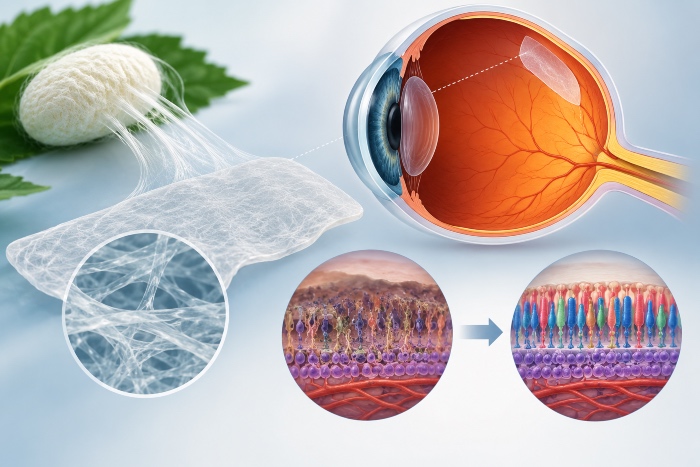
The retinal pigment epithelium is a monolayer of strongly polarized cuboidal cells, interposed between the outer segments of the photoreceptors and the choriocapillaris and anchored on its basal side to Bruch's membrane. From this position it governs a set of processes indispensable to vision, from the daily phagocytosis of shed outer-segment discs to the recycling of retinoids that fuels the visual cycle, from the vectorial transport of ions, water and metabolites between the neural retina and the choroidal circulation to the absorption of stray light mediated by melanin, through to the polarized secretion of trophic factors, with PEDF directed apically toward the photoreceptors and VEGF released on the basolateral side to sustain the integrity of the choriocapillaris. The failure of this layer is the event that triggers or accompanies age-related macular degeneration, both in the atrophic form with geographic atrophy and in the exudative form with choroidal neovascularization, and it characterizes numerous inherited retinal dystrophies in which the genetic defect is expressed primarily in the RPE itself.
Why injecting cells in suspension is not enough
The earliest regenerative strategies relied on the subretinal injection of cell suspensions, yet this approach runs into biological limits that are hard to circumvent. RPE cells are anchorage-dependent epithelial cells and, once deprived of a substrate on which to spread, they undergo anoikis, lose their apico-basal polarity and fail to reconstitute the tight junctions that define the outer blood-retinal barrier. To this is added the fact that, in the degenerated eye, Bruch's membrane is itself altered, thickened and laden with deposits, and offers an inadequate bed for engraftment. What follows is a strong rationale for delivering an already formed, mature and polarized monolayer, supported by a prosthetic Bruch's membrane that acts at once as a surgical carrier and as a provisional matrix. It is in this space that silk fibroin has established itself as one of the most extensively studied biomaterials.
The properties that make fibroin a prosthetic Bruch's membrane
Fibroin satisfies a profile of requirements that few materials meet simultaneously. It is biocompatible and weakly immunogenic once the sericin has been removed, it exhibits slow and tunable enzymatic degradation, it tolerates processing carried out entirely in the aqueous phase and, not least, it can be rendered optically transparent, a feature that is far from irrelevant for an implant destined to sit along the visual axis. Its chemistry moreover allows stiffness, porosity and resorption kinetics to be regulated independently, making it possible to decouple parameters that in other systems remain bound together. The comparison with the alternatives clarifies the advantage. Synthetic polyesters such as PLGA release acidic degradation products that can acidify the subretinal microenvironment, amniotic membrane suffers from marked donor-dependent variability, and collagen films tend toward poorly controllable degradation. Fibroin occupies a favourable intermediate position, combining the reliability of a natural polymer with a predictability of behaviour close to that of synthetic materials.
Structure and processing of regenerated fibroin
The behaviour of the finished membrane is decided upstream, in the chemistry of the protein and in its processing. Bombyx mori fibroin is organized into a heavy chain and a light chain joined by a disulphide bridge, with hydrophobic domains that tend to pack into beta sheets. The degree of crystallinity in the silk II configuration, as opposed to the amorphous silk I form, is the main lever governing both mechanical strength and degradation rate. The production of regenerated fibroin begins with degumming, which removes the sericin responsible for most of the inflammatory responses, continues with dissolution in lithium bromide and dialysis, and concludes with shaping. In thin-film substrates, the addition of poly(ethylene oxide) as a porogen introduces controlled porosity, while treatment with methanol or water-vapour annealing induces the transition to beta sheets and stabilizes the membrane by modulating its stability. The versatility of the available formats completes the picture, from solvent-cast films to nanofibrous membranes obtained by electrospinning, through to hydrogels. A further option is offered by the wild fibroin of Antheraea pernyi, which unlike that of Bombyx mori natively contains RGD motifs capable of promoting integrin-mediated cell adhesion without the need for exogenous functionalization.
The biomimetic constraints imposed by the native Bruch's membrane
Every design choice has to reckon with the architecture of the membrane it intends to replace. The native Bruch's membrane is a pentalaminar lamina of just two to four micrometres, which thickens with age and which must ensure the bidirectional passage of nutrients, catabolites and fluids between RPE and choroid. A scaffold destined for the subretinal space must therefore be ultrathin, because exceeding the tolerable thickness means compromising its placement and disturbing its relationship with the overlying photoreceptors; some lenticular carriers, with thicknesses on the order of tens of micrometres, have shown precisely this limitation. The Bombyx mori fibroin membranes developed as a Bruch's membrane substitute have been obtained with thicknesses of around three micrometres, permeability coefficients between roughly three and nine times ten to the minus five centimetres per second, and pores on the order of a few micrometres, values compatible with the metabolic exchange required in situ. Mechanical compliance, which must approach that of the host tissue, remains central as well, together with degradation kinetics ideally synchronized with the deposition of endogenous extracellular matrix by the transplanted cells, so that resorption of the support proceeds in step with the construction of its biological replacement.
Adhesion, polarization and maturation of the RPE on the substrate
Geometric compatibility alone is not sufficient, because the RPE expresses its own phenotype only if the substrate guides its adhesion and polarization. On fibroin membranes, optimal attachment of the ARPE-19 line has been achieved by pre-coating the surface with vitronectin at concentrations on the order of one microgram per millilitre, and analogous strategies employ laminin, type IV collagen or RGD peptides to engage the cellular integrins. When the dialogue between cell and matrix is correct, the cultures develop the cobblestone morphology typical of the mature epithelium, with cortical distribution of F-actin, tight junctions marked by ZO-1, apical microvilli and basal infoldings, accompanied by the appearance of pigmentation. Maturation is documented at the molecular level through the expression of markers such as RPE65 and CRALBP of the visual cycle, bestrophin encoded by BEST1, the transcription factor MITF and the melanosome proteins, and at the functional level through the transepithelial electrical resistance that indexes barrier tightness, the polarized secretion of PEDF and VEGF, and the capacity to phagocytose photoreceptor outer segments. It is significant that RPE cultures maintained for weeks on nanofibrous fibroin substrates have preserved precisely the polarized secretion of PEDF and phagocytic activity, two of the functions hardest to preserve in vitro and among the most predictive of physiological behaviour after implantation.
From the cell line to pluripotent stem cells
Much of the early characterization relied on the ARPE-19 line, useful as a model but unsuited to clinical use because of its incomplete phenotype. The decisive step was to demonstrate that fibroin membranes support the maturation of RPE derived from stem cells, first embryonic and then induced pluripotent. The generation of a functional RPE monolayer from hiPSC on a prosthetic Bruch's membrane made of Bombyx mori fibroin represented in this sense a landmark result, because it linked a well-characterized biomaterial to a scalable and clinically viable cell source. On the translational level the choice of source is not neutral. Autologous iPSC eliminate the problem of histocompatibility and reduce the recourse to immunosuppression, but they impose individual production times and costs hardly compatible with broad diffusion, whereas allogeneic lines selected for HLA compatibility and organized into cell banks promise an off-the-shelf product, at the price of more careful management of the immune response, all the more so since the immune privilege of the retina presupposes an intact RPE and Bruch's membrane, precisely the condition that has failed in the patient to be treated.
In vivo evidence and clinical perspective
The in vivo profile of fibroin in the ocular setting is encouraging. Composite nanofibrous membranes of wild fibroin, polycaprolactone and gelatin, produced by electrospinning with thicknesses of three to five micrometres and fibres of nanometric diameter, have supported cultures of human RPE for up to twelve weeks and, implanted subsclerally in animal models, have proved biocompatible with no sign of inflammatory reaction. The position of fibroin within the clinical landscape must nonetheless be stated honestly. The RPE-on-scaffold implants that have reached human trials have so far relied above all on polyester and parylene membranes carrying embryonic-stem-cell-derived RPE, on PLGA patches with iPSC-derived RPE, on amniotic membrane and on fibrin hydrogels, while fibroin remains today a candidate at the advanced preclinical stage. Its combination of transparency, tunable degradation, aqueous processing and reproducible behaviour nonetheless makes it one of the natural materials with the greatest likelihood of clinical translation, in a field where the choice of carrier bears on the outcome as much as the quality of the transplanted cells.
