
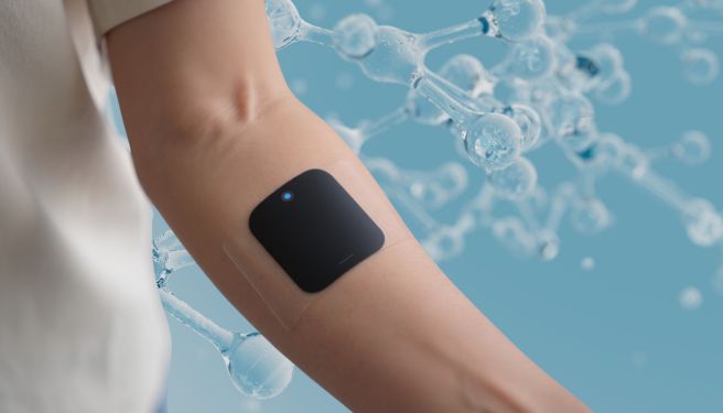
The concept of a cutaneous bioscreen — a display conformal to the skin surface, flexible, biocompatible, and integrated with sensing functions — represents the point of convergence between fibroin research and epidermal electronics technologies. The main technical challenge is that skin is not a flat and rigid surface: it deforms, sweats, changes its surface impedance depending on hydration, and poorly tolerates rigid or occlusive materials in prolonged contact.
Fibroin modified with calcium chloride (CaCl?) develops a viscoelastic behavior that allows the film to conform to the micro-geometries of the skin, including grooves, pores, and papillary ridges, without generating concentrated stress areas. Ca²? ions act on two levels: on one hand they plasticize the protein structure by reducing stiffness, on the other they chelate the random-coil chains, creating adhesion points that generate interfacial cohesion and energy dissipation during cyclic deformation. Four different types of epidermal electronics have been demonstrated to adhere to human skin through this silk hydrogel, all operating under normal skin-use conditions.
The flexible display based on composite fibroin and AgNWs has been proposed for applications on human skin by exploiting the fact that any body segment — forearm, back of the hand, wrist — can act as a display substrate. Silk fibroin ionic touchscreen technology (SFITS) recently described in the literature introduces an additional functional level: through a humidity-induced crystallization strategy, the molecular structure of fibroin is precisely controlled to obtain a balance between mechanical robustness, ionic conductivity, and biodegradability. The device has been shown to operate under different environmental conditions and integrated with IoT functionalities and artificial neural networks for physiological signal classification.
In parallel, fibroin-MXene devices represent a hybrid approach in which two-dimensional MXene nanosheets (transition-metal carbides and nitrides) are incorporated into the protein matrix to provide high conductivity and multimodal sensing capability, including pressure, temperature, and strain detection — all variables that a cutaneous bioscreen with diagnostic functionality must acquire simultaneously with visualization.
Programmable biodegradability and transient electronics
One of the most extraordinary technical properties of fibroin for implantable biomedical devices is its programmable biodegradability — not the generic, random one of synthetic biodegradable polymers such as PLA or PGA, but an enzymatic degradation that can be controlled through the β-sheet crystallinity of the material.
In vivo degradation of fibroin is mediated by tissue proteases (mainly serine proteases and matrix metalloproteinases), and the degradation rate is inversely proportional to the β-sheet content: highly crystalline films degrade over weeks or months, more amorphous structures over hours or days. This means that engineering the secondary structure of the film determines not only the optical and mechanical properties but also the operational time window of the device before its controlled biological dissolution. The degradation products — amino acids and short peptides — are not only non-toxic but are metabolized and reabsorbed by the host tissue without accumulation, eliminating the need for surgical removal.
In transient fibroin devices with AgNWs, substrate dissolution occurred in 1M NaOH on the order of minutes, while under physiologically similar conditions (protease solution) the process extended to weeks, corresponding to biologically relevant times for a temporary implantable diagnostic device. The degradation timescale can be tuned during fabrication by acting on the annealing process (methanol annealing vs water annealing vs thermal annealing), which determines the crystallization pattern of the film.
For transient cutaneous displays, dissolution of the protein substrate in sweat or body humidity is a limitation that must be engineered: the ionic composition of sweat (pH 4.5–7.5, NaCl 20–100 mM, presence of lactate and glucose) is in some cases sufficient to plasticize less stabilized fibroin films. The technological response consists in the use of the previously described doping strategies (PU, IPA, CaCl?) to obtain the required stability window without compromising long-term degradability.
Nonlinear optical properties
Beyond display applications, recent characterizations of the nonlinear optical properties of fibroin open technically relevant perspectives for next-generation implantable photonic systems. Measurements using the Z-scan technique with femtosecond laser pulses (35 fs, 800 nm, 1 kHz) revealed a strong self-defocusing effect (negative nonlinear refractive index) and significant multiphoton absorption in thin fibroin films. The nonlinear refractive index n? of fibroin exceeds that of fused quartz by one order of magnitude, while the multiphoton absorption coefficient β is about two orders of magnitude higher.
These data position fibroin as a candidate for second-harmonic generation (SHG) and optical frequency conversion applications, functions that in implantable photonics could allow optical information transmission through biological tissue at wavelengths different from the excitation one — opening the way to sub-dermal optical communication systems between implantable devices and external readers.
The photoelasticity of fibroin, combined with its nonlinear optical properties and its ability to be nanopatterned through soft lithography or protein-protein imprinting (PPi), enables the realization of reconfigurable optical elements: resonant microcavities whose resonance wavelength changes in response to mechanical deformation of the substrate — a mechano-optical sensing principle that can be integrated into a biomedical display to encode diagnostic information in color or light-intensity variations, without the need for additional active electronics.
Development directions
The path toward fully functional fibroin-based implantable biomedical displays and cutaneous bioscreens still faces several concrete technical barriers. The first is the management of the fibroin-AgNW interface in physiodynamic environments: silver nanowires tend to oxidize in biological environments rich in ionic chlorine, with a consequent increase in contact resistance and potential release of cytotoxic Ag? ions at high concentrations. Studied solutions include protective coating of the nanowires with ultrathin SiO? or TiO? layers via ALD, or their replacement with functionalized carbon-nanotube networks or GrapheneMesh, both chemically more stable.
The second concerns the scalability and uniformity of large-area fibroin films. Solution casting from aqueous systems produces films with local thickness variations linked to solvent-evaporation gradients, which translate into refractive-index and transmittance non-uniformities on the centimeter scale — incompatible with high-resolution display specifications.
Dynamic coating techniques (slot-die coating, doctor blading on temperature-controlled heated substrates) and spin-coating from formic-acid solution — a benign solvent that produces flatter and more uniform films than water or HFIP — have been demonstrated as partial solutions, but industrial-scale process standardization remains an unmet goal.
The third is wireless power delivery to the implantable display: an e-ink system does not require continuous power to maintain the image, but the state transition is still energetically costly, and NFC coils for inductive energy transfer through tissue must be integrated without adding unacceptable mechanical rigidity. Genetic engineering of silkworms — including emerging CRISPR modifications to optimize batch-to-batch the amino-acid composition of produced fibroin — could in the medium term provide proteins with optical and mechanical characteristics even more finely engineered than what is currently achievable through post-extraction chemical processing.
