
Rheological and mechanical properties for neuro-mimicry
In the context of advanced neural interfaces, the ability to replicate the mechanical properties of nervous tissue represents a crucial design requirement. Brain parenchyma exhibits an extremely low elastic modulus, typically ranging between 0.1 and 10 kPa under physiological conditions, and any significant mechanical mismatch is known to trigger inflammatory responses and undesired cellular remodeling. In this framework, fibroin stands out for its structural versatility: its supramolecular organization into hydrophobic β-sheet domains alternating with hydrophilic amorphous regions allows fine modulation of elastic properties through control of crystallinity degree and hydration state. Rather than intrinsically matching nervous tissue, fibroin can be engineered to approach such values, reducing mechanical mismatch compared to traditional rigid substrates such as silicon or metal oxides. Several studies suggest that low-stiffness substrates contribute to limiting the activation of mechanosensitive signaling pathways, including the RhoA/ROCK axis, with favorable effects on neuronal morphology and the stability of dendritic arborizations. In vivo models, fibroin-based devices generally display an attenuated inflammatory response compared to conventional materials, with reduced astrogliosis and glial scar formation, though without a complete absence of glial reactivity.
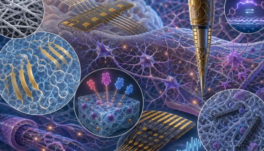
Design of composite conductive scaffolds
An intrinsic limitation of fibroin is its electrically insulating nature, which restricts its direct use in electrophysiological devices. To overcome this constraint, composites are developed by integrating nanostructured conductive fillers. The incorporation of multi-walled carbon nanotubes (MWCNTs), typically functionalized to improve their dispersion, enables the formation of percolative networks even at low concentrations (on the order of 1% by volume). These systems exhibit a significant reduction in electrochemical impedance — on the order of tens of kΩ at 1 kHz, depending on electrode geometry — and a charge injection capacity compatible with safe neural stimulation applications. Alternatively, integration with conductive polymers such as PEDOT:PSS yields hybrid materials with mixed ionic-electronic conduction. In this context, fibroin plays a dual role: a mechanically compliant matrix and a hydrophilic phase that facilitates ionic transport, contributing to reduced interfacial impedance compared to traditional metal electrodes. Conductivity values can vary widely depending on the doping level and processing conditions, reaching, in optimized configurations, tens of S/cm.
Guided topographies and neurite outgrowth
Beyond physicochemical properties, micro- and nanotopography plays a decisive role in the interaction with neural tissue. Techniques such as soft lithography on elastomeric molds or electrospinning make it possible to generate anisotropic architectures capable of guiding neuronal growth. Aligned fibrous scaffolds, with diameters on the order of hundreds of nanometers and micrometric spacing, promote axonal extension along preferential directions through contact guidance mechanisms. In primary cultures, such structures support extensive axonal growth (up to the millimeter scale over timescales of days) and a high degree of alignment. For penetrating applications, fibroin can be processed into three-dimensional microstructures, including microneedles. In these devices, the combination of a flexible core and conductive coatings (such as gold and iridium oxide) allows adequate electrochemical properties to be maintained while reducing the mechanical damage associated with tissue micro-movements.
Controlled release of neurotrophins and biohumoral integration
Processability in aqueous environments allows the direct incorporation of biomolecules during fabrication. Neurotrophic factors such as NGF or BDNF can be loaded into the matrix with good encapsulation efficiencies, varying as a function of the gelation method and physicochemical conditions. Release typically follows anomalous diffusion kinetics, with an initial burst phase followed by prolonged delivery over time. Although near zero-order profiles have been observed under specific conditions, in most cases release is better described as sustained rather than perfectly constant. Fibroin degradation is tunable through β-sheet content: treatments with solvents such as methanol increase the crystalline fraction, slowing biodegradation from a few weeks to several months. During this process, peptides derived from the protein matrix are released, which may contribute to cell adhesion and the migration of supportive glial cells such as Schwann cells.
The integration of photosensitive components opens the way to multifunctional neural interfaces. Photoisomerizable molecules, such as azobenzene derivatives, can induce reversible changes in mechanical properties and ionic permeability under light stimulation, although the extent of these effects depends strongly on the system's chemistry and degree of functionalization. In parallel, the incorporation of fluorescent probes or nanoparticles enables optical monitoring of the tissue-device interface, including visualization of phenomena such as the glial response. Emerging applications include the localized delivery of viral vectors for optogenetics, a field still in an exploratory phase but promising for the spatially controlled modulation of neuronal activity. The good optical transparency of fibroin, particularly at low crystallinity levels, also makes it suitable for devices requiring coupling between light stimulation and electrical recording.
In preclinical models of nervous system injury, fibroin-based scaffolds have demonstrated the ability to support axonal regrowth and to variably improve functional outcomes compared to controls. These effects are generally associated with a reduction in inhibitory regeneration signals and better organization of newly formed tissue. In cortical applications, flexible devices based on fibroin and conductive polymers prove capable of recording neural signals with quality comparable to traditional materials, but with a more contained tissue response in the medium-to-long term. It is important to emphasize that, while results are promising, there is significant variability across studies in terms of protocols, animal models, and evaluation metrics.
