
Silk fibroin is experiencing a second youth in the contemporary bioelectronics landscape, transforming from a simple structural protein into a sophisticated technological platform for applications requiring electrical conductivity in biological environments. This functional metamorphosis is based on the ability to chemically and physically modify fibroin to confer electrical properties while maintaining intact the characteristics that have made it so interesting for medicine: exceptional biocompatibility, controllable biodegradability, tunable mechanical properties, and processability into different forms. Conductive fibroin today represents a meeting point between materials science, biomedical engineering, and neuroscience, opening application scenarios that until a few years ago seemed relegated to science fiction.
The concept of making fibroin conductive arises from the need to create bioelectronic devices that can intimately interface with biological tissues without provoking adverse long-term reactions. Traditional conductive materials used in medical settings, such as metals and rigid synthetic polymers, present significant problems related to the difference in mechanical impedance with soft tissues, the generation of chronic inflammatory responses, and difficulty in biological integration. Fibroin modified to become conductive instead offers an alternative that combines adequate electrical performance with a practically ideal biological interface, capable of integrating with the surrounding tissue environment without creating that "barrier" which characterizes conventional devices.
Functionalization strategies for conductivity
The methodologies for conferring conductive properties to fibroin are multiple, and each presents specific advantages depending on the final application. The most widespread approach involves the incorporation of conductive nanomaterials within the protein matrix, creating hybrid composites in which fibroin provides the biocompatible structure and the conductive material ensures charge transport. Carbon nanotubes, graphene, and its derivatives such as reduced graphene oxide represent the most common choices for this strategy. These carbonaceous materials are dispersed in the aqueous fibroin solution before its gelation or polymerization, generating percolative networks that guarantee conductivity even at relatively low concentrations. The key to success lies in obtaining a homogeneous distribution of conductive nanomaterials and optimizing their concentration: too few and conductivity remains inadequate, too many and the mechanical properties of fibroin are compromised.
An elegant alternative is represented by the incorporation of organic conductive polymers such as polyaniline, polypyrrole, or PEDOT:PSS. These materials, being also polymeric, show better compatibility with the protein matrix compared to rigid carbonaceous nanomaterials. The strategy generally involves in situ polymerization of the conductive monomer within preformed fibroin structures, or direct blending of the two polymers in solution. PEDOT:PSS in particular has gained considerable attention thanks to its stability in aqueous environments, high conductivity, and processability. The presence of the sulfonic group in PSS also favors electrostatic interactions with the amino groups of fibroin, improving dispersion and composite stability. These hybrid materials can reach conductivities in the order of 10?² - 10?¹ S/cm, values sufficient for most bioelectronic applications.
Then there are more sophisticated approaches that involve surface metallization of fibroin structures through deposition of ultra-thin metallic films, typically gold or silver, or through bioinspired mineralization processes that lead to controlled growth of metallic nanoparticles on the protein surface. These techniques allow obtaining very high conductivities while maintaining the bulk of the material in biocompatible fibroin, creating a stratified architecture where electronics interface with the surface metal while biological tissue interacts mainly with the underlying fibroin.
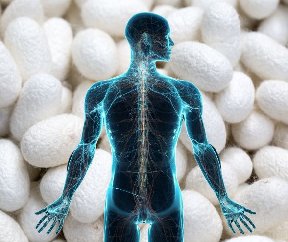
Neurostimulation: when electrical signals speak to the nervous system
The application of conductive fibroin in neurostimulation probably represents the most promising and challenging field. Electrical neurostimulation is an established therapeutic technique that uses controlled electrical impulses to modulate neuronal activity, finding use in the treatment of neurological conditions such as Parkinson's disease, drug-resistant epilepsy, chronic pain, and movement disorders. The conventional electrodes used in these devices are however made of rigid metallic materials which, despite their effectiveness in charge transfer, present significant problems when chronically implanted in nervous tissue. The difference in elastic modulus between metal and nervous tissue (of several orders of magnitude) generates micromovements that cause chronic inflammation, formation of scar tissue (gliosis), and progressive loss of interface functionality. This is where conductive fibroin can make a difference.
Electrodes made with composites of fibroin and conductive materials offer mechanical properties much closer to those of nervous tissue, drastically reducing mechanical stress at the interface. This mechanical compatibility translates into a reduction in inflammatory response and prolonged maintenance of signal quality over time. Recent studies have demonstrated that flexible fibroin-graphene based electrodes implanted in the brain of animal models maintain stable and low impedances for periods exceeding six months, against the progressive deterioration observed with rigid metallic electrodes under the same conditions. The surface of fibroin, moreover, can be functionalized with neuroactive peptides or growth factors that promote neuronal integration, transforming the electrode from foreign body to biologically integrated component of nervous tissue.
The geometry of conductive fibroin electrodes can be optimized for specific neurostimulation applications. For deep brain stimulation (DBS), arrays of ultra-flexible microelectrodes have been developed that can conform to the microtopography of brain tissue, minimizing damage during insertion. For spinal cord stimulation in the treatment of chronic pain, conductive fibroin patches have been created that adhere intimately to the dura mater surface, distributing stimulation over larger areas with greater uniformity compared to point electrode arrays. The ability of fibroin to be processed through microfabrication techniques such as soft lithography, stamping, and electrospinning also allows creating complex electrode patterns with high spatial resolution, opening the way to high-density neural interfaces capable of recording and stimulating with single-neuron precision.
Electrotherapy and muscle stimulation: from laboratory to clinic
Electrotherapy represents another field in which conductive fibroin is demonstrating remarkable potential. This therapeutic modality uses electrical currents to stimulate muscles, improve circulation, reduce pain, and accelerate healing processes. Conventional devices use adhesive surface electrodes which, while effective, present limitations in terms of duration, contact stability, and possibility of inducing skin irritation with prolonged use. Conductive fibroin allows overcoming these limits through the creation of biointegrated textile electrodes that can be comfortably worn for extended periods.
Conductive fabrics based on fibroin are produced through electrospinning of solutions containing fibroin and conductive materials, creating nanofibers that are then assembled into three-dimensional matrices. These fibrous structures show excellent breathability, conformability to the skin surface, and uniformly distributed conductivity, characteristics that make them ideal for wearable electrotherapy applications. Unlike commercial electrodes that concentrate current in specific points creating unpleasant or even painful sensations, conductive fibroin fabrics distribute stimulation over larger surfaces, making the experience much more comfortable for the patient. This characteristic is particularly important in applications requiring prolonged daily treatments, such as functional electrical stimulation in patients with spinal cord injuries.
Functional electrical stimulation (FES) deserves particular attention. This technique uses electrical impulses to artificially activate paralyzed or weakened muscles, restoring compromised motor functions. Conductive fibroin electrodes for FES can be surgically implanted in direct contact with the muscle belly or motor nerves, offering a much more stable and durable interface compared to conventional electrodes. The superior biocompatibility of fibroin reduces the formation of fibrous scar tissue around the electrode, maintaining low electrical impedance and guaranteeing efficient charge transfer over time. Some research groups are developing completely implantable FES systems based on fibroin-graphene electrodes connected to flexible electronic circuits, creating completely biodegradable devices designed to support the initial rehabilitation phase after neurological injuries and then gradually dissolve once functional recovery has been achieved.
Neurological rehabilitation: accelerating functional recovery
The field of neurological rehabilitation is experiencing a radical transformation thanks to the integration of advanced bioelectronic technologies, and conductive fibroin is playing an increasingly important role in this evolution. Rehabilitation after stroke, spinal injuries, or cranial trauma requires the reactivation of damaged neural circuits and the formation of new synaptic connections, processes that can be significantly accelerated through controlled electrical stimulation. Rehabilitation devices based on conductive fibroin offer unique advantages in this context, combining the ability to provide therapeutic electrical stimulation with properties that actively support tissue regeneration.
Three-dimensional scaffolds in conductive fibroin represent a particularly interesting platform for peripheral nervous tissue regeneration. When a nerve is severed, axon regeneration through the lesional gap requires a physical substrate that guides neuritic growth. Neural conduits made with fibroin-polypyrrole composites simultaneously provide a mechanical template for axonal growth and the possibility to apply electrical stimulation which, as demonstrated by numerous studies, significantly accelerates the regenerative process. The conductivity of the material allows applying weak but continuous electrical fields that orient axon growth in the desired direction (phenomenon known as galvanotaxis), while the porous microarchitecture of the scaffold provides spaces for Schwann cell migration, essential for myelination of new axons. Preclinical studies have shown that the use of these conductive conduits can reduce nerve regeneration times by thirty to forty percent compared to non-conductive conduits, with superior functional recovery.
In the context of central nervous system rehabilitation, conductive fibroin devices are finding application as interfaces for cortical recording and stimulation during neurorehabilitation assisted by brain-computer interfaces. These systems read the patient's brain electrical activity and use it to control external devices (such as exoskeletons or prosthetic limbs) or to provide stimulative feedback to the brain itself, creating neuroplasticity loops that accelerate functional recovery. Epicortical electrode arrays made in fibroin-graphene offer superior signal quality thanks to their ability to perfectly conform to the irregular cortical surface, reducing movement artifacts and improving signal-to-noise ratio. Their flexibility also allows less invasive implantation and greater duration compared to conventional rigid arrays.
Bioelectronic interfaces: beyond stimulation
Bioelectronic interfaces represent the most advanced frontier in the application of conductive fibroin, pushing the concept of medical device toward that of bio-synthetic hybrid system capable of bidirectional communication with biological tissues. Unlike simple stimulating electrodes, advanced bioelectronic interfaces can both record endogenous bioelectrical signals (such as neuronal action potentials or electromyographic potentials) and provide controlled stimulation in response to these signals, creating intelligent closed-loop systems. Conductive fibroin lends itself particularly well to this type of application thanks to the combination of adequate electrical conductivity, optical transparency (which allows imaging techniques through the device), permeability to nutrients and metabolites, and ability to be functionalized with active biological elements.
Three-dimensional bioelectronic interfaces made with conductive fibroin hydrogels represent a particularly sophisticated evolution. These materials, typically obtained through incorporation of PEDOT:PSS or graphene in fibroin hydrogels, combine the mechanical properties of soft tissues (elastic modulus in the order of kPa) with electrical conductivity in the order of 10?³ - 10?² S/cm. This combination allows the creation of porous scaffolds that cells can colonize three-dimensionally, generating electrically active engineered tissue constructs. Neurons, cardiomyocytes, or skeletal muscle cells grown in these scaffolds maintain their electroactivity and can be stimulated or recorded through the material itself, without the need for traditional metallic electrodes. This technology is opening fascinating possibilities in the field of electrically functional organoids and in vitro models of excitable tissues for drug screening.
The ability to integrate flexible electronics directly onto conductive fibroin structures has led to the development of multimodal implantable biosensors that can simultaneously monitor electrophysiological, biochemical, and mechanical parameters. These devices use conductive fibroin as a biocompatible substrate on which to deposit ultra-thin electronic circuits, electrochemical sensors for detection of neurotransmitters or metabolites, and piezoelectric transducers for mechanical sensing. The integration of these multiple functionalities into a single thin and flexible device allows much more complete monitoring of the physiological state of tissue compared to traditional devices that measure only electrical parameters. In the context of closed-loop neurostimulation, for example, these devices could not only detect pathological neuronal activity but also monitor local levels of neurotransmitters and the metabolic response of tissue, allowing much finer and more personalized therapeutic control.
Technical challenges and future perspectives
Despite remarkable progress, the clinical implementation of conductive fibroin still faces significant challenges that require further technological developments. Long-term stability of conductivity in physiological environment represents an important concern, particularly for composites based on organic conductive polymers that can degrade in the presence of reactive oxygen species generated by inflammatory processes. Delamination of interfaces between conductive materials and fibroin can lead to loss of electrical properties over time, requiring chemical functionalization strategies that guarantee stable covalent bonds between components. Carbonaceous nanomaterials, while offering greater chemical stability, pose questions regarding their biodegradability and long-term metabolic fate, aspects still not completely clarified.
The scalability of production processes represents another important challenge. Many of the techniques used to incorporate conductive materials into fibroin are labor-intensive and difficult to standardize, making the transition from academic research to industrial production problematic. The development of robust, reproducible manufacturing protocols compatible with regulations for medical devices is essential for clinical translation. Sterilization of finished devices without compromising the properties of conductive materials represents an aspect not to be underestimated, since many conductive polymers are sensitive to the high temperatures of autoclaving and to solvents used in other sterilization methods.
From a regulatory point of view, conductive fibroin finds itself in a gray zone between traditional biomaterials and active electronic devices, requiring approval pathways that consider both the biocompatibility of the protein matrix and the safety and efficacy of the conductive component. The intrinsic variability of fibroin extracted from natural sources can also pose standardization problems that regulatory agencies require for approval of medical devices. The use of recombinant fibroin produced in heterologous systems could offer a solution to this problem, guaranteeing batches of material with reproducible molecular characteristics.
Technological convergence and personalized medicine
Looking to the future, the true revolution of conductive fibroin might not reside so much in individual applications as in the convergence of multiple technologies that this material platform makes possible. The integration of biosensing, adaptive stimulation, controlled drug delivery, and regenerative scaffolding in multifunctional devices represents a particularly promising direction. Imagine implantable devices that not only stimulate nervous tissue but also monitor neurochemical markers of inflammation and release anti-inflammatory agents in response, all while gradually degrading once the therapeutic process is completed. Conductive fibroin, with its versatility of processing and functionalization, could be the ideal material to realize this vision.
Additive manufacturing and biofabrication techniques are opening unprecedented customization possibilities. The ability to 3D print complex conductive fibroin structures directly based on the patient's specific anatomy, derived from medical imaging, would allow creating bioelectronic interfaces perfectly adapted to individual morphology, maximizing therapeutic efficacy and minimizing side effects. This precision medicine extended to the level of material-tissue interface represents a paradigm shift compared to current "one-size-fits-all" devices.
Integration with artificial intelligence technologies for analysis of bioelectrical signals and real-time optimization of stimulation parameters constitutes another fascinating frontier. Machine learning algorithms could learn from patterns of neuronal or muscular activity recorded through conductive fibroin interfaces, automatically adapting stimulation strategies to maximize therapeutic benefit for each patient. This combination of intelligent materials and intelligent software could lead to truly autonomous neuromodulation systems, capable of operating effectively with minimal medical supervision.
Conductive fibroin is thus establishing itself not only as an innovative material but as an enabling technological platform for a new generation of bioelectronic therapies. Its unique ability to mediate between the world of electronics and that of biology, providing an interface that speaks both languages fluently, positions it at the center of a revolution that is redefining the boundaries between medical devices, engineered tissues, and biological organisms. The path from basic research to widespread clinical application is still long, but the scientific and technological foundations are solid and rapidly expanding, revealing a future in which the distinction between natural and artificial becomes increasingly blurred, and in which materials are no longer inert supports but active participants in healing and regeneration processes.
