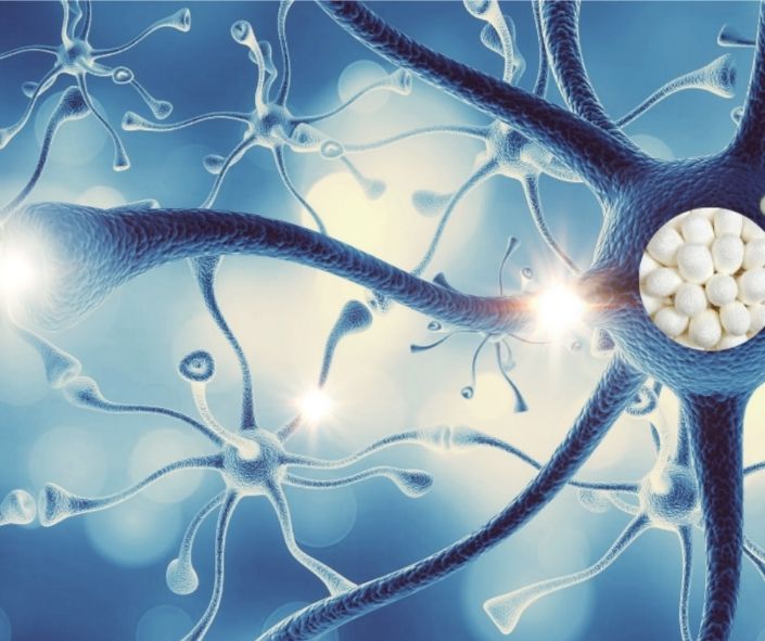
The process of transforming fibroin into a conductive material requires sophisticated functionalization strategies that preserve biocompatibility while conferring electroactive properties. The most advanced research has demonstrated that the integration of conductive nanomaterials such as reduced graphene, carbon nanotubes, and conductive polymers like polypyrrole can transform fibroin scaffolds into effective electroactive platforms. Studies have shown how coating electrospun fibroin fibers with reduced graphene creates materials that combine excellent electrical conductivity with the ability to support neuronal growth.
The most widespread technique involves electrospinning fibroin to create nanofibers that mimic the natural extracellular structure of nervous tissues, followed by surface coating or also by direct incorporation of conductive materials. In the case of reduced graphene, for example, deposition often occurs through controlled chemical processes that allow obtaining a uniform layer on the surface of fibroin fibers. This configuration maintains the nanostructured morphology essential for guiding axon growth, while the conductive component allows the passage of low-intensity electrical currents that stimulate regenerative processes.
A particularly interesting alternative approach involves the use of two-dimensional materials such as MXenes, a class of metallic carbides and nitrides with extraordinary electrical conductivity and hydrophilicity properties. The combination of fibroin with MXenes has produced injectable hydrogels that can be implanted directly at the site of nerve injury, creating a conductive microenvironment that promotes the differentiation of neural stem cells into functional neurons. These systems represent a significant step forward compared to rigid scaffolds, as they can adapt to the complex geometries of damaged tissues and integrate perfectly with the surrounding tissue architecture.
Neurostimulation through conductive fibroin scaffolds for peripheral and central nervous system regeneration
Nervous system injuries are a complex reality in medicine, as adult neurons have extremely limited regenerative capacities, especially in the central nervous system. In this context, conductive scaffolds based on fibroin have extraordinary potential in facilitating nerve regeneration through multiple mechanisms that go beyond simple structural support. The presence of conductive properties indeed allows applying controlled electrical stimulations that mimic the endogenous signals of the nervous system, accelerating axonal growth and improving the myelination of regenerating nerve fibers.
Studies conducted on spinal cord injury models in rats have shown how aligned scaffolds of graphene and fibroin can guide the directional growth of neurites, reproducing the fascicular architecture of the original nerve tracts. The alignment of fibers is not just a geometric factor, but also creates preferential conductive pathways that facilitate the propagation of electrical signals along the regeneration axis. This spatial organization is crucial because growing neurons naturally follow the directions imposed by support structures, and the presence of conductivity along these pathways further enhances the guiding effect.
Regarding the peripheral nervous system, where regenerative capacities are greater but often insufficient for complete functional recovery, the use of nerve conduits made of conductive fibroin has shown promising results. These tubular devices can be designed with multichannel architectures that replicate the fascicular structure of peripheral nerves, and their conductive component allows applying electrical fields that orient and accelerate axonal growth across the lesion gap. The combination with neurotrophic growth factors encapsulated in the fibroin matrix creates a synergistic environment where chemical and electrical stimuli cooperate to maximize functional recovery.
Implantable bioelectronic interfaces
Beyond three-dimensional scaffolds for regeneration, fibroin also finds crucial applications in the creation of implantable bioelectronic interfaces that allow recording and modulating the electrical activity of nervous and muscular tissues. These devices, often realized as thin films with incorporated electrode patterns, represent an elegant solution to the long-term biocompatibility problem that afflicts conventional metallic electrodes. Fibroin, in fact, is not only tolerated by the organism for prolonged periods, but also tends to integrate with surrounding tissues, reducing the formation of scar tissue that typically degrades the performance of electronic implants.
The typical structure of these devices includes a flexible fibroin substrate on which conductive electrodes are deposited using lithography or printing techniques. The sandwich configuration, with electrodes incorporated between two layers of fibroin, protects the conductive components from the biological environment while maintaining a completely biocompatible external interface. This architecture is particularly advantageous for applications on the cerebral cortex or peripheral tissues, where the mechanical conformability of the device is essential to prevent mechanical damage due to relative movements between the implant and the host tissue.
Recent studies have demonstrated that these devices maintain high sensitivity in detecting electromyographic and electroencephalographic signals for periods exceeding one week in vivo, without evidence of significant performance degradation. This temporal stability is fundamental for clinical applications where continuous monitoring of neural activity is necessary for diagnosis or to implement adaptive therapeutic strategies. Furthermore, the optical transparency of fibroin allows combining electrical recordings with optical imaging techniques, creating multimodal platforms for studying neural activity with superior spatial and temporal resolution.
Muscle electrotherapy mediated by conductive fibroin biomaterials
In the context of muscle rehabilitation, functional electrical stimulation represents an established strategy to prevent atrophy, promote strength recovery, and facilitate motor relearning after neurological injuries and direct muscle trauma. However, conventional surface electrodes also present significant limitations in terms of stimulation selectivity, patient comfort, and long-term therapeutic adherence. The integration of conductive materials based on fibroin in wearable and implantable devices thus presents itself as an innovative solution to overcome these limitations, offering more conformable, biocompatible interfaces capable of more targeted stimulations.
Fibroin scaffolds functionalized with conductive polymers such as polypyrrole have been studied specifically for cardiac and skeletal muscle tissue engineering applications. These materials allow not only supporting the growth and alignment of muscle cells in vitro, but also applying electrical fields that synchronize cellular contractions and promote the functional maturation of engineered tissue. In the rehabilitation context, this technology could translate into bioengineered muscle patches capable of integrating with native muscle and responding to electrical stimulations to facilitate recovery after volumetric muscle injuries.
A particular application concerns closed-loop systems that combine electromyographic monitoring with adaptive electrical stimulation. In these systems, sensors made with conductive fibroin detect the residual electrical activity of the damaged muscle and use this information to modulate in real time the intensity and timing of the applied electrical stimulation. This approach has been successfully experimented in volumetric muscle injury models, where robot-assisted stimulation has enabled immediate recovery of gait through selective activation of lower limb muscles when electromyographic activity exceeded certain thresholds.
The intrinsic piezoelectric properties of fibroin and their exploitation for cellular stimulation
A less known but extremely fascinating aspect of fibroin concerns its intrinsic piezoelectric properties, which allow the protein to generate electrical potentials in response to mechanical deformations. This characteristic, deriving from the structural organization of protein chains in beta-sheet conformation, opens completely new possibilities for electrical stimulation of nerve and muscle cells without the need for external energy sources or complex electronic circuits. The principle is relatively simple: when fibroin nanofibers are deformed by physiological movements of the surrounding tissue or by mechanical forces applied during rehabilitation, they generate electrical microcurrents that can influence cellular behavior.
Many studies have focused on the structural optimization of fibroin to maximize the piezoelectric response, intervening on the crystallinity of the material and on the molecular orientation of protein chains. It has been demonstrated that highly crystalline nanofibers, obtained through mechanical stretching treatments or also through immersion in specific solvents, show significantly higher piezoelectric coefficients compared to amorphous forms of the same protein. This possibility of modulating piezoelectric properties through material processing offers considerable control over the final characteristics of the device.
The application of piezoelectric fibroin scaffolds in nerve regeneration has shown encouraging results, with evidence that cyclic mechanical stimulation of these materials promotes neurite elongation and accelerates neuronal differentiation even in the absence of direct external electrical stimulation. This mechanism is particularly interesting for in vivo applications where the patient's simple motor activity could provide the necessary mechanical stimulation to generate beneficial electrical signals, creating a self-sustained stimulation system that does not require batteries or implanted electronic components.
Enhanced neurological rehabilitation through fibroin conductive materials
In the field of advanced neurological rehabilitation, materials based on conductive fibroin can play a key role as a biocompatible interface both for recording brain signals and for applying stimulation to paralyzed limbs. The operational paradigm provides that the patient imagines or attempts to perform a specific movement, the associated brain activity is recorded through fibroin electrodes positioned on the motor cortex or on the scalp, and these signals are decoded in real time to command electrical stimulation of target muscles through electrodes also made with biocompatible conductive materials.
The superiority of fibroin in these systems mainly resides in its ability to maintain stable, high-quality interfaces with biological tissues for prolonged periods. Conventional electrodes in metal or synthetic polymers tend to induce chronic inflammatory responses that progressively degrade the quality of the recorded signal and the effectiveness of the applied stimulation. Conversely, electrodes incorporated in fibroin matrices show superior tissue integration, with minimal formation of glial scar tissue in the case of cortical implants, and greater stability of electrical impedance over time.
Experimental applications of these integrated systems have demonstrated significant improvements in the recovery of upper limb motor function in post-stroke patients when functional electrical stimulation is synchronized with motor intention detected from brain activity. This approach exploits residual neural plasticity, since the temporal association between voluntary cortical activity and proprioceptive feedback generated by electrically induced muscle contraction reinforces damaged neural circuits and promotes functional reorganization of cerebral motor areas. The use of conductive fibroin materials at both ends of this neuro-rehabilitative circuit ensures that the biological interface remains optimal during the entire treatment period, which typically extends for weeks or even for months.
Technological challenges in electrochemical and mechanical stabilization of fibroin-conductive material composites
Research is focusing on stabilization strategies that include chemical cross-linking between fibroin and conductive materials, the incorporation of antioxidant protective agents, and optimization of the biological component-conductive component ratio to balance electrical performance and durability. A promising approach involves the use of ultra-thin protective coatings of pure fibroin over conductive layers, creating a fluid-permeable barrier capable of significantly slowing degradative processes. This configuration maintains surface biocompatibility while protecting the underlying conductive infrastructure.
From a mechanical point of view, the decoupling of elastic properties between naturally flexible fibroin and some rigid conductive materials such as carbon nanotubes can generate interfacial tensions that lead to detachment or fracture of components under cyclic stress. The solutions explored include the use of intrinsically flexible conductive materials such as MXenes or ultra-thin metallic nanowires that can deform together with the fibroin matrix, or the implementation of serpentine architectures for electrodes that allow large deformations without accumulation of mechanical stress. These engineering considerations are fundamental to ensure that devices maintain their functionality during physiological movements, particularly critical for muscular or articular implants subject to continuous cycles of extension and contraction.
Future perspectives and translational potential of conductive fibroin bioelectronic systems
Looking to the future, the evolution of conductive biomaterials based on fibroin is orienting towards increasingly sophisticated systems that integrate sensory, actuating, and computational capabilities in completely biocompatible platforms. The ultimate goal is to create fully implantable bioelectronic devices that can continuously monitor the functional state of nervous and muscular tissues, provide adaptive therapeutic stimulations in response to detected signals, and communicate wirelessly with external systems for medical control and adjustment of therapeutic parameters. Fibroin, with its unique combination of biocompatibility, processability, and functionalization capability, positions itself ideally as a material platform to realize this vision.
A particularly interesting area of development concerns the incorporation of miniaturized electronic circuitry directly into fibroin matrices, creating hybrid systems where biological and synthetic components coexist in an integrated architecture. Recent progress in the fabrication of biocompatible transistors and sensors allows imagining fibroin scaffolds that not only passively conduct electrical signals, but can also amplify, filter, and process them locally before transmission. This distributed computational capability would significantly reduce the energy requirements and complexity of implantable devices, bringing closer the realization of completely autonomous neural prostheses.
From the perspective of personalized medicine, the ease with which fibroin can be processed into complex forms through three-dimensional printing techniques or computerized electrospinning opens the possibility of creating custom-made devices based on the specific anatomy of the patient obtained from medical imaging. A patient with peripheral nerve injury could receive a conductive nerve conduit designed exactly to bridge the gap in their particular anatomy, with optimized distribution of conductive materials and growth factors based on computational models of nerve regeneration. This personalization would also extend to electrical stimulation parameters, which could be individually calibrated based on the electrophysiological response measured in real time through the same fibroin electrodes.
Perhaps the most revolutionary aspect concerns the potential controlled biodegradability of these systems. While conventional electronic implants require secondary surgical interventions for removal once the therapeutic function is completed, conductive fibroin devices could be designed to progressively degrade as the regenerated tissue acquires autonomous functionality. The degradation kinetics could be modulated through chemical modifications of fibroin or through device geometry, ideally synchronizing the disappearance of artificial support with the completion of natural regeneration. This strategy would eliminate the risks associated with long-term presence of foreign bodies and would represent a paradigm shift toward truly transient bioelectronic interventions that leave behind only functional regenerated tissue.


