
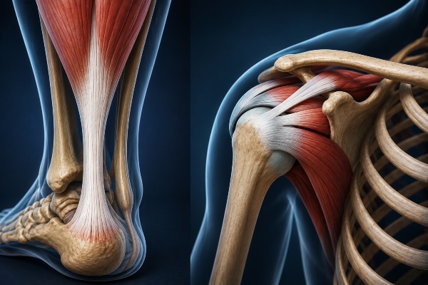
The Achilles tendon and the rotator cuff represent two of the most demanding districts in the entire field of tissue engineering, not so much because of the biological complexity of the tendon itself as because of the mechanical environment in which any attempt at repair must prove functional. An implant that regenerates correctly in culture but fails at the first physiological stress has no clinical value, and it is precisely here that silk fibroin finds its reason for being. The structural protein produced by Bombyx mori possesses a combination of tensile strength, toughness and controlled degradation that few other natural matrices manage to offer, and the way it behaves under cyclic loading makes it a particularly serious candidate for tendon reconstruction.
The mechanical problem of tendon healing
The tendon is a dense, poorly vascularized tissue organized into parallel bundles of type I collagen that align along the axis of force application. This hierarchical architecture, descending from fibrils to fibers to fascicles, is what allows the tendon to efficiently transmit forces even ten times greater than body weight. When the tissue is injured, spontaneous repair almost always produces disorganized scar tissue, rich in type III collagen, mechanically inferior and prone to new ruptures. In the Achilles tendon this translates into a concrete risk of re-rupture after treatment, while in the rotator cuff failure typically manifests at the tendon-to-bone interface, where the suture tends to tear through the degenerated tissue before biology can even intervene.
The engineering challenge therefore consists not only in providing a scaffold on which cells can deposit new matrix, but in ensuring that this scaffold withstands physiological load throughout the entire time required for maturation of the neotissue. A material that is too rigid ends up shielding the cells from the mechanical stimuli they need, a phenomenon known as stress shielding, while a material that is too weak fails before regeneration is complete. The useful window is narrow, and fibroin fits into it because it is possible to modulate both its initial mechanical properties and its degradation kinetics.
Why fibroin responds well to cyclic loading
Fibroin's strength arises from its secondary structure. The crystalline regions organized into antiparallel beta sheets, rich in repeated sequences of glycine, alanine and serine, confer on the material remarkable rigidity and tensile strength, while the interposed amorphous regions absorb energy and restore elasticity. This alternation between rigid domains and yielding domains reproduces, on a molecular scale, the same principle of energy dissipation that makes biological tendon so tough. It is one of the reasons why fibroin scaffolds do not merely resist static load but tolerate repeated stress well, which is exactly the regime to which a tendon is exposed during walking, running or elevation of the arm.
Behavior under mechanical fatigue is the true testing ground. Numerous studies have shown that fibroin constructs retain a good part of their tensile properties after thousands of loading and unloading cycles, a result that many synthetic polymeric matrices reproduce only at the cost of an acidic degradation unfavorable to tissues. Fibroin, degrading enzymatically through proteases, releases peptides and amino acids that the body reabsorbs without generating the inflammatory microenvironment typical of polyesters such as polyglycolic acid. This balance between mechanical retention and biocompatible degradation is precisely what is needed at a site subjected to permanent load.
From scaffold shape to function
Perhaps fibroin's most relevant advantage is its processing versatility, because the same protein can be transformed into very different geometries according to the biomechanical requirement. For tendon reconstruction the most promising strategy exploits fibrous, aligned structures, obtained by electrospinning or by weaving filaments, capable of guiding the orientation of tendon cells along the load axis. Topographical alignment is not an aesthetic detail, because tenocytes cultured on oriented substrates assume an elongated morphology and deposit collagen along the direction of the fibers, reproducing the anisotropy that makes native tendon so efficient in transmitting force.
Fibroin-based scaffolds for tendon use are often built as hierarchical structures, in which thin fibers assemble into thicker bundles that mimic the multilevel organization of the real tendon. This design makes it possible to calibrate the overall elastic modulus so that it approaches that of the host tissue, reducing the mechanical mismatch at the interface with the suture and distributing the load more uniformly. In the rotator cuff, where the critical point is the enthesis, the complex gradual transition between tendon, fibrocartilage and bone, gradient fibroin scaffolds are being explored, with increasing mineralization toward the bony end, so as to reconstruct not only the tendon but also the anchoring zone that ensures its hold.
The dialogue between material and cells
A mechanically sound matrix is not enough if it does not support the biological response. Fibroin offers surfaces that favor cell adhesion and can be functionalized with recognition sequences such as the RGD motif, with growth factors or with other extracellular matrix proteins to direct the differentiation of stem cells toward the tenogenic phenotype. In this sense the scaffold becomes an active element, capable not only of bearing the load but of transmitting to the cells the mechanical signals that induce them to behave like tenocytes. The load itself, applied in a controlled manner through mechanical stimulation protocols in a bioreactor, significantly improves the quality of the tissue produced, because cyclic tension promotes the alignment of newly formed collagen fibers and increases their strength.
It is here that the most interesting synergy between fibroin and the mechanical environment emerges. Instead of considering load as a threat from which the implant must be protected, the most advanced approach uses it as a therapeutic stimulus, entrusting to fibroin the task of bearing it in the initial phase and transmitting it to the cells in physiological doses as the neotissue matures and progressively takes on responsibility for the load. This gradual transfer of function, from the synthetic material to the biological tissue, is the true objective of modern tendon repair, and the timed degradation of fibroin makes it achievable.
