
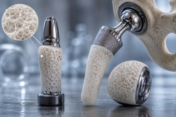
For decades, prosthetic orthopedics has lived with a paradox that materials science has learned to recognize but not yet to fully resolve. A hip or knee prosthesis, or an internal fixation device, must integrate stably with the host bone, establishing a mechanical and biological bond meant to last for years; at the very same moment, however, the implant surface represents a territory ripe for conquest by microorganisms, an inert platform on which bacteria can settle before the host's own cells have a chance to colonize it. This is the celebrated race for the surface described by Anthony Gristina in the late 1980s: a silent contest between the tissue that seeks to adhere and the biofilm that seeks to proliferate, in which whoever arrives first dictates the fate of the implant. Silk fibroin, is one of the few biomaterials capable of acting simultaneously on both fronts of this race, offering coatings that promote osseointegration while at the same time hindering bacterial colonization.
Why the implant surface is the real battleground
Titanium alloys, and Ti-6Al-4V in particular, remain the gold standard of prosthetic orthopedics owing to their excellent strength-to-weight ratio and a biocompatibility that time has confirmed. Titanium, however, is a bioinert material: its spontaneous oxidation forms a stable, chemically quiet TiO? layer that does not actively dialogue with bone cells. Osseointegration — that direct, functional contact between living bone and the implant surface first described by Per-Ingvar Brånemark — therefore occurs despite the metal's inertness, not because of it. When integration is incomplete or delayed, the result is a weak interface, micromotion, fibrous encapsulation and, ultimately, aseptic loosening, one of the leading causes of revision surgery.
To this biological fragility is added the risk of infection. Periprosthetic infections, the so-called prosthetic joint infections, are a formidable complication not so much for their absolute incidence, which remains contained, as for the difficulty of treating them. Staphylococci such as Staphylococcus aureus and Staphylococcus epidermidis adhere to the implant surface and organize themselves into a biofilm, a sessile community embedded in an extracellular matrix of exopolysaccharides that acts as a shield. Within the biofilm, bacteria slow their metabolism, evade immune defenses and tolerate antibiotic concentrations even a hundred or a thousand times higher than those effective against planktonic forms. Once established, a biofilm rarely regresses with systemic therapy alone, and treatment often requires surgical removal of the implant, with all that this entails in terms of morbidity, cost and patient quality of life.
The problem, in other words, cannot be solved by choosing between sound bony anchorage and effective antimicrobial protection: both must be achieved, on the same surface, in the same critical window of the first hours and days after implantation. And it is precisely here that an intelligent coating — rather than a merely passive one — makes the difference.
Silk fibroin as a biomaterial platform
Fibroin is the structural component of the silk fiber, distinct from sericin, the gluey protein that coats it and that is removed during the degumming process. At the molecular level, fibroin owes its properties to a repetitive sequence dominated by glycine, alanine and serine, organized into hydrophobic domains that pack into antiparallel β-sheets. It is precisely these crystalline β-sheet regions that confer on the material its remarkable mechanical strength and its stability in a physiological environment, while the amorphous regions provide elasticity and toughness. The ability to modulate the ratio between the crystalline and amorphous phases — through treatments such as exposure to methanol, ethanol, water vapor or thermal processing — makes it possible to govern precisely the degradation rate and the properties of the coating, tailoring them to the requirements of the anatomical site.
What makes fibroin especially suited to coating orthopedic implants is a combination of features that rarely coexist in a single material. It is biocompatible and elicits a modest inflammatory response, lower than that of many synthetic polymers. It is biodegradable through physiological enzymatic pathways, with non-toxic amino acid degradation products, and its resorption kinetics can be extended from weeks to months or years. It possesses robust mechanical properties that allow it to adhere firmly to a metallic substrate and to withstand the stresses of surgical insertion. Above all, it is extraordinarily versatile from a processing standpoint: it can be handled entirely in the aqueous phase, at room temperature and under mild conditions, which makes it possible to incorporate fragile bioactive molecules — growth factors, antibiotics, peptides — without denaturing them. This "gentle chemistry" is the reason fibroin works so well as a reservoir and as a vehicle for controlled release.
How silk fibroin promotes osseointegration
The first task of an osseointegrative coating is to transform a mute metallic surface into a substrate that cells recognize and on which they choose to settle. Fibroin fulfills this role by offering osteoblasts a protein-rich environment to which they can adhere, proliferate and differentiate. The surface of a silk film, when suitably structured, supports cell adhesion and the deposition of extracellular matrix, and several in vitro studies have documented increased expression of osteogenic markers such as alkaline phosphatase, osteocalcin and bone sialoprotein on coated surfaces compared with bare titanium.
The real leap in quality, however, comes with functionalization. Fibroin lends itself to combination with hydroxyapatite, the calcium phosphate that constitutes the mineral phase of bone, generating composite coatings in which the protein component provides the organic scaffold and the mineral component offers nucleation sites for mineralization. These organic–inorganic coatings reproduce in miniature the very nature of bone tissue, which is itself a nanocomposite of collagen and apatite, and they accelerate the formation of new bone at the interface. In some strategies, hydroxyapatite is precipitated directly onto the fibroin film by incubation in simulated body fluid, exploiting the protein's carboxyl groups as nucleation centers and thereby achieving a biomimetic mineralization that grows from within the coating rather than being merely deposited on top of it.
Alongside mineralization, fibroin can serve as a reservoir for signaling molecules that orchestrate bone regeneration. Incorporating growth factors such as BMP-2, the bone morphogenetic protein that drives the differentiation of mesenchymal cells along the osteoblastic lineage, makes it possible to transform the coating from a passive substrate into an active inducer of osteogenesis. The β-sheet structure protects the growth factor from degradation and modulates its release, avoiding the burst release that would needlessly disperse its activity. Likewise, functionalization with adhesion peptides bearing the RGD sequence strengthens the bond between cellular integrins and the coating, improving osteoblast anchorage in the early phases. Topography, too, plays no minor role: the nano- and microstructure of the fibroin film, controllable at the deposition stage, influences cell orientation and behavior, and an appropriate roughness stimulates the osteogenic response better than a smooth surface does.
The antibacterial strategies of silk coatings
If osseointegration is the promise, infection is the threat, and a modern coating must guard against both. Fibroin intervenes on the antimicrobial front chiefly by virtue of its ability to act as a matrix for the controlled release of bactericidal agents, but also, to a more modest degree, through surface modifications that hinder initial bacterial adhesion.
The most direct and most widely studied approach is the loading of antibiotics. Vancomycin, gentamicin, ciprofloxacin and other molecules can be incorporated into the fibroin film during its aqueous-phase formation, becoming trapped within the protein network. Their subsequent release is governed by the microstructure of the coating and by its degree of crystallinity: increasing the fraction of β-sheets slows diffusion and prolongs the therapeutic window, yielding a high local antibiotic concentration exactly where it is needed — at the bone–implant interface — without exposing the entire organism to systemic effects. This local, sustained release is particularly valuable in the first hours after surgery, the window in which the race for the surface is decided, and it makes it possible to strike at bacteria before they organize a mature biofilm.
A complementary strategy, independent of antibiotics and therefore less exposed to the problem of antibiotic resistance, is the incorporation of metallic nanoparticles, silver above all. Silver exerts broad-spectrum bactericidal action through the release of Ag? ions that disrupt bacterial membranes, interfere with replication and generate reactive oxygen species. Fibroin has proven an excellent matrix for the synthesis and stabilization of silver nanoparticles, which can be generated in situ by reducing precursors directly within the protein, achieving a homogeneous distribution and a controlled ionic release that balances antibacterial efficacy against cellular tolerability. The same approach has been explored with nanoparticles of copper, zinc oxide and other inorganic systems, each with its own activity profile.
More recent and promising is the use of antimicrobial peptides, short cationic amino acid sequences that destabilize bacterial membranes and that, unlike conventional antibiotics, rarely induce resistance. The chemical compatibility between these peptides and fibroin — both being protein entities workable in an aqueous environment — makes silk a natural vehicle for their presentation and gradual release. There are also approaches that aim not to kill bacteria but to prevent their adhesion, rendering the surface antifouling through chemical modification of the film or the grafting of hydrophilic chains, so as to deny microorganisms their first foothold.
A coating that does both
The frontier of research lies not in producing coatings that are either antibacterial or osseointegrative, but in combining the two within a single architecture without allowing the two functions to obstruct one another. This balance is anything but a given, because many antimicrobial agents — silver in particular — are cytotoxic at high concentrations and may compromise the very osteogenic cells one is trying to favor. Designing a bifunctional coating therefore requires careful calibration of the dose and release kinetics of the bactericidal agent, so that the window of maximum antimicrobial activity coincides with the period of greatest infection risk and then yields to the osteoconductive component as bony integration gains momentum.
The most elegant solutions exploit fibroin's structural versatility to organize the coating into compartments or gradients. A layered architecture — built, for instance, with the layer-by-layer technique, alternating silk strata loaded with antibiotic or silver with strata mineralized with hydroxyapatite — makes it possible to program a temporal sequence: first the release of the antibacterial agent from the outermost layers, then the progressive exposure of the osteoconductive surface as the surface layers degrade. In this way the same coating "changes its job" over time, defending the surface during the critical phase and then handing it over to the host's cells. It is the material, almost chronobiological, translation of the race for the surface: not winning one of the two contests, but winning both in the right order.
How fibroin coatings are deposited onto metal
Translating these promises into a clinically useful device depends largely on the technique by which the fibroin is applied to the substrate. The available methods differ in thickness, uniformity, adhesion and ability to coat complex geometries, and the choice depends on the type of implant and the intended function.
Dip coating and spin coating are the simplest and most widely used methods at the laboratory scale: the former immerses the implant in the fibroin solution, yielding thin and relatively uniform films even on irregular surfaces, while the latter distributes the solution by centrifugation and is ideal for flat substrates. Electrospinning, by contrast, generates nanofibrous membranes that reproduce the architecture of the extracellular matrix, offering a high specific surface area and a topography that cells favor, particularly suited to maximizing cellular interaction. Electrophoretic deposition harnesses an electric field to migrate and deposit fibroin — optionally co-precipitated with hydroxyapatite — onto conductive substrates such as titanium, ensuring good adhesion and control over thickness. The layer-by-layer technique already mentioned, finally, builds the coating by alternating assembly of oppositely charged layers, and it is the royal road to multifunctional architectures with sequential release.
A recurring practical issue is the adhesion of the protein film to the metallic surface, which can be improved through preliminary treatments of the titanium such as plasma activation, anodic oxidation or silane functionalization, capable of creating a stable chemical anchorage between metal and protein. The robustness of this bond is decisive, because a coating that delaminates during surgical insertion not only loses its function but may also generate harmful debris.
