
Closing a wound remains one of the oldest gestures in medicine, and for millennia it had only one truly reliable instrument: thread. Sutures and staples still work admirably when the surgeon operates in an open field with room to maneuver, yet they reveal their limits precisely where contemporary surgery is moving with the greatest determination, namely inside millimetric accesses, through trocars, catheters and endoscopes, on soft, friable or richly vascularized tissues that tolerate the traction of a stitch poorly. Every perforation introduces an additional trauma, every knot demands manual precision and operative time, and in many anatomical sites mechanical hold is not enough, because what is really needed is a continuous seal against the loss of air, blood or fluids. It is in this space, between the need to hold the edges together and the need to seal them, that bioadhesive systems have ceased to be a marginal accessory and have become an enabling technology of minimally invasive surgery. And in this very space silk fibroin is emerging as one of the most interesting raw materials on which to build the next generation of surgical glues and sealants.
Why adhesives have not yet replaced thread
Tissue adhesives have existed for decades, and yet none of those available today has won the role of universal standard, the reason being that each one pays a price. Cyanoacrylates polymerize within a few seconds and offer a robust grip, but they release potentially cytotoxic by-products during degradation and tend to form a rigid, brittle film that is poorly tolerated on dynamic internal tissues. Fibrin glues reproduce the last step of the coagulation cascade and are excellent for hemostasis and biocompatibility, yet they adhere weakly, resorb quickly, derive from blood products with their attendant costs and theoretical risk of transmission, and rarely withstand high pressures. Polyethylene glycol-based sealants guarantee good hold but swell in situ, and that swelling can become a problem in confined spaces such as the spinal canal. There also remains a difficulty common to nearly all of these families, that is adhesion in a wet environment: blood and biological fluids interpose a hydrated veil between the adhesive and the tissue surface, and on that veil most chemistries lose their efficacy. A material that truly aspires to replace thread must therefore reconcile requirements in tension with one another, adhering strongly to wet tissue without damaging it, remaining flexible enough to accompany the motion of a lung or a loop of intestine, resorbing on predictable timescales and demonstrating an impeccable biocompatibility. It is a demanding profile, and it is precisely by reading this list of requirements that fibroin enters the scene.
Fibroin as a molecular platform
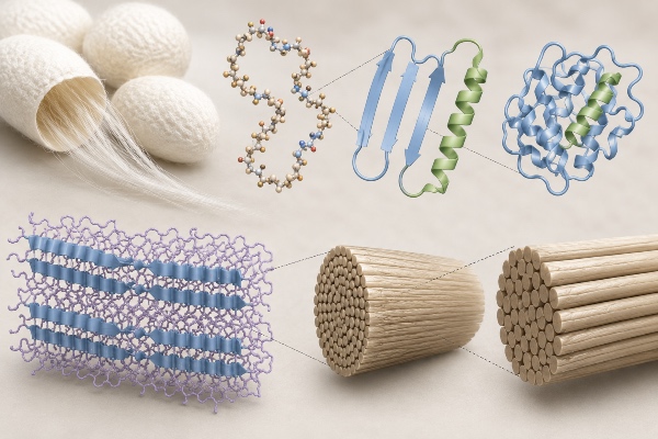
Fibroin is the structural protein that constitutes the filamentous core of the cocoon of the silkworm Bombyx mori, stripped of the sericin that coats it. Its architecture is that of a heavy chain of roughly 390 kilodaltons joined by a disulfide bridge to a light chain of about 26 kilodaltons, accompanied by an accessory glycoprotein. What makes it mechanically extraordinary is the alternation, along the heavy chain, of crystalline hydrophobic blocks rich in repeated glycine-alanine-glycine-alanine-glycine-serine motifs, and of more hydrophilic amorphous regions. Those repeated blocks have a tendency to stack into antiparallel beta-sheets stabilized by a dense network of hydrogen bonds, and it is precisely these crystalline domains that endow silk with the otherwise rare combination of tensile strength and toughness. The protein regenerated in the laboratory can be induced to pass from the metastable conformation, sometimes designated silk I, to the stable beta-sheet form, silk II, and this transition is the lever by which the stiffness, stability and, above all, degradation rate of the final material are controlled.
What makes fibroin so appealing for surgery is not a single property but their convergence. It is a protein that is notoriously biocompatible and poorly immunogenic, processable entirely in water and at room temperature, a condition that allows drugs and biological factors to be incorporated without denaturing them. It degrades enzymatically into metabolizable amino acids over time windows that range from weeks to months depending on the crystallinity that is set, avoiding both the overly rapid resorption that penalizes fibrin and the indefinite persistence of certain synthetic polymers. It is available in large quantity and at low cost from a consolidated agricultural supply chain, and it can take practically any useful form in the operating room, from a thin film to an injectable gel, from an expandable sponge to a sprayable solution. Only one piece is missing from this almost ideal picture, and it is also the most delicate.
The knot of adhesion in a wet environment
Native fibroin, in itself, is not a good glue for wet tissues. Its internally cohesive crystalline regions give the material its solidity, but they offer few chemical functionalities capable of actively binding to a biological surface, and in the presence of water the interaction tends to remain superficial. To transform an excellent structural protein into an effective surgical adhesive it is therefore necessary to introduce, chemically or physically, the reactive groups that nature did not give it in abundance. The good news is that fibroin lends itself surprisingly versatilely to being functionalized, and that each of the strategies developed in recent years answers to a different logic of adhesion, often inspired by solutions that biology has already road-tested.
The path that has attracted the most attention is the mussel-mimetic one. Mussels adhere firmly to submerged rocks thanks to proteins rich in a catecholic amino acid, DOPA, whose catechol groups form covalent and coordination bonds with a wide range of surfaces, including wet and mineralized ones. By conjugating fibroin with dopamine, or coating it with polydopamine, this anchoring capacity is transferred to it: the catechols react with the amino and thiol groups of tissue proteins and establish stable interactions even across the film of water, resolving precisely the problem that throws traditional adhesives into crisis. A second strategy instead exploits an amino acid that fibroin already possesses, tyrosine, present in sufficient measure to be put to use. By oxidizing the tyrosine residues, di-tyrosine bonds form that crosslink the protein network and, at the same time, can latch onto the tyrosines exposed on the tissue. This oxidation can be triggered enzymatically, typically with horseradish peroxidase in the presence of hydrogen peroxide or with tyrosinase, obtaining hydrogels that gel within a few seconds, or photochemically, employing photoactivatable ruthenium complexes that under visible light generate the same bonds without requiring aggressive reagents.
Alongside these two main lines, the toolbox has grown richer. The methacrylation of fibroin produces a photo-crosslinkable version, commonly called silk methacrylate, which in the presence of a photoinitiator solidifies in a rapid and spatially controlled manner under irradiation, a feature that is precious when one wants the adhesive to harden only where and when it is needed. Crosslinking with genipin, a natural compound that binds primary amino groups, offers a low-toxicity alternative to synthetic crosslinkers. And blending with polyphenols such as tannic acid, or with polysaccharides such as chitosan, adds further hydrogen-bonding and coordination sites that enhance both internal cohesion and adhesion to the substrate. The common direction of all these routes is the same: to preserve the structural qualities of fibroin and graft onto it a surface chemistry that makes it capable of gripping living tissue.
Injectable formulations and in situ gelation
Minimally invasive surgery imposes on the adhesive a constraint that does not exist in the open field, namely that of deliverability. A material that is excellent but rigid, to be applied by hand on an exposed surface, is useless if it must pass through a trocar a few millimeters wide or the working channel of an endoscope to reach a deep site. Hence the interest in injectable formulations that remain fluid while passing through the instrument and gel once they reach their destination, with kinetics slow enough to allow positioning yet rapid enough not to disperse into the surrounding fluids.
The enzymatic di-tyrosine chemistry has proved particularly suited to this purpose, because it allows a fibroin solution to be injected that crosslinks in situ within seconds when the oxidation is activated, forming a cohesive, adhesive gel exactly where it has been deposited. The photo-crosslinkable versions offer an even finer control, because gelation occurs only at the moment the surgeon illuminates the area, leaving full freedom of maneuver up to that point. There are also formulations in the form of sponges or shape-memory matrices, compressed for passage through the miniaturized access and capable of re-expanding on contact with fluids, filling irregular cavities and adapting to the geometry of the defect. In all these cases fibroin's workability in water at room temperature is a decisive advantage, because it avoids organic solvents and heat, both poorly tolerated in tissues and incompatible with any incorporated active ingredients.
Sealants, films and patches for hold
The objective is not always to glue two edges together: often what counts is to seal. Sutures leave microspaces between one stitch and the next, and in many situations the feared complication is not mechanical dehiscence but leakage through those microspaces, whether it be air from a pulmonary suture, cerebrospinal fluid from a dural breach, bile from a biliary anastomosis or enteric content from an intestinal suture. Here fibroin lends itself to being shaped into conformable films and thin patches that, applied over the suture line, create a continuous barrier. The parameter that measures the value of these devices is the burst pressure, that is the maximum pressure the seal withstands before yielding, and the most advanced fibroin adhesive formulations have shown, in preclinical models, values competitive with commercial standards while maintaining at the same time a flexibility that the rigid film of cyanoacrylates does not possess.
Fibroin's capacity to take on thin, transparent geometries also makes it suitable for contexts where conformability to the irregular, moving surface of an organ matters. A patch that accompanies the expansion of a lung without detaching, or that follows the peristalsis of an intestinal loop without cracking, reduces the risk that the seal will give way in the postoperative period, when the tissue resumes its physiological dynamics. The ability to regulate the degree of crystallinity during fabrication makes it possible to calibrate the rigidity and duration of the barrier according to the site, leaving a device that holds as long as needed and then resorbs as healing proceeds.
Hemostasis and tissue repair
An ideal surgical sealant does not merely hold in place what it encounters: it actively contributes to stopping bleeding and to healing the wound. On the hemostatic front fibroin works on several levels. As a porous matrix, in its sponge or foam versions, it provides an extended surface on which platelets adhere and become activated and on which the clot organizes itself, while at the same time offering a physical tamponade. To this structural action can be added the loading of procoagulant agents, obtaining devices that combine mechanical hold with the direct promotion of hemostasis, a precious combination in operative fields difficult to control by compression alone.
On the regenerative front, fibroin brings with it a quality that few adhesives possess, that of being itself a good substrate for cells. Fibroin surfaces support the adhesion, proliferation and activity of numerous cell populations, modulate the inflammatory response favorably, and degrade leaving room for newly formed tissue rather than obstructing it. An adhesive built on this protein, therefore, is not merely a glue destined to disappear, but can behave like a temporary scaffold that guides repair, aligning its own resorption rate with the timing of tissue regeneration. This dual nature, sealant and regenerative at once, is probably the trait that most sharply distinguishes fibroin from purely mechanical alternatives.
Integrated functions: drug release and antibacterial activity
The fully aqueous, low-temperature processing of fibroin opens a possibility that goes beyond simple adhesion, namely transforming the sealant into a therapeutic vehicle. Antibiotics, antimicrobial peptides, growth factors and metal nanoparticles can be incorporated into the matrix during fabrication and then released in a sustained manner as the material degrades, with kinetics that are once again tunable by adjusting the crystallinity. A patch that seals an anastomosis and simultaneously releases an antibiotic at the surgical site addresses, in a single gesture, two distinct risks, leakage and infection, and a sealant that frees growth factors can accelerate the repair of the tissue on which it is applied. It is in this convergence of functions that fibroin perhaps shows its most ambitious promise, because it shifts the bioadhesive from the role of a passive component to that of a local therapeutic device, capable of acting where the operation has left its mark.
