
Sericin exhibits osteoinductive properties through molecular mechanisms that actively stimulate the formation of new bone tissue. This capability is expressed through the modulation of specific cellular pathways that guide progenitor cells toward the osteoblastic phenotype, accelerating bone mineralization and remodeling processes. Scientific evidence accumulated in recent years positions sericin as a molecule of particular interest for orthopedic and dental applications, where bone regeneration represents a crucial clinical challenge.
Mechanisms stimulating osteoblastic differentiation
Sericin directly intervenes in the processes that transform mesenchymal stem cells into mature osteoblasts, the cells responsible for synthesizing the bone matrix. Research conducted on human mesenchymal cell cultures has demonstrated that sericin significantly increases the expression of key osteogenic markers such as Runx2, the master transcription factor of osteogenesis, and osterix, which is essential for osteoblastic maturation. These transcription factors orchestrate the activation of hundreds of genes involved in bone formation, triggering a complex genetic program that leads to cellular differentiation.
The osteoinductive effect of sericin is manifested through activation of the Wnt/β-catenin signaling pathway, a fundamental pathway in the regulation of osteogenesis. Studies published in Biomaterials have shown that sericin promotes the stabilization and nuclear translocation of β-catenin, enabling the activation of target genes such as those encoding alkaline phosphatase, osteocalcin, and type I collagen. Alkaline phosphatase represents a crucial enzyme in the early stages of mineralization, while osteocalcin constitutes the main non-collagenous protein of the bone matrix, binding calcium and facilitating the formation of hydroxyapatite crystals.
Sericin also modulates the BMP/Smad pathway, considered one of the most powerful signaling cascades in osteogenic induction. Bone morphogenetic proteins (BMPs) are growth factors that stimulate bone formation, and sericin enhances the cellular response to these signals. Experiments have shown that mesenchymal cells cultured on sericin-containing substrates display increased phosphorylation of Smad1/5/8 proteins, intracellular mediators of BMP signaling, with a consequent increase in the expression of osteogenic genes. This synergistic effect between sericin and endogenous or exogenous BMPs suggests particularly effective therapeutic combinations.
Modulation of the extracellular environment for mineralization
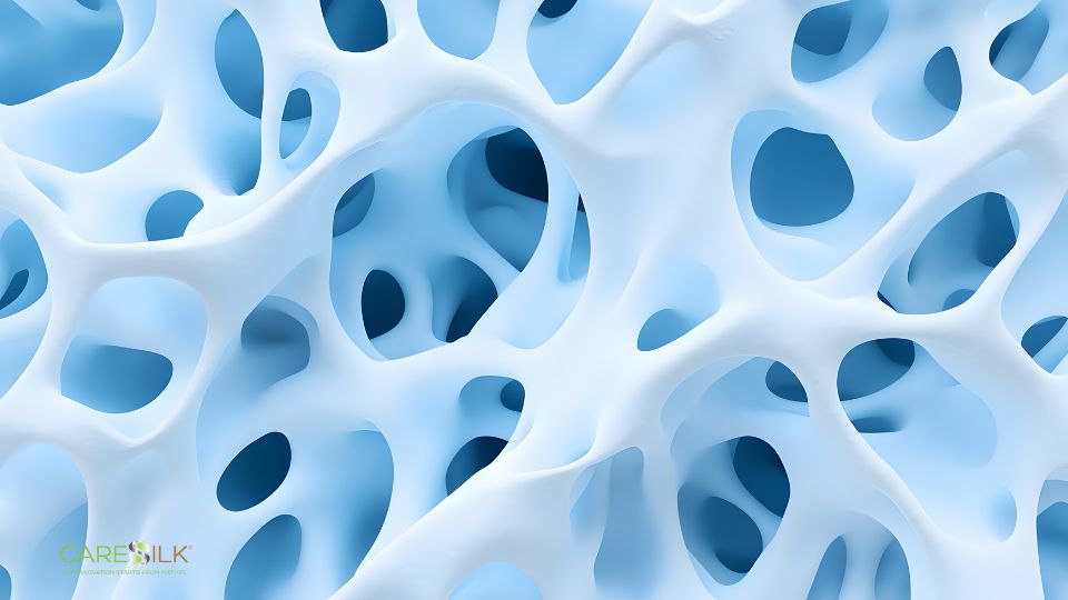
The ability of silk protein to influence the composition and organization of the extracellular matrix contributes significantly to its osteoinductive properties. Sericin promotes the deposition of type I collagen, the main organic component of bone, creating a molecular scaffold suitable for subsequent mineralization. Electron microscopy analyses have revealed that the presence of sericin favors the organization of collagen fibrils into more ordered and aligned patterns, optimizing the matrix structure for the incorporation of mineral crystals.
Sericin directly interacts with calcium phosphate crystal nucleation processes, facilitating the formation of hydroxyapatite, the main mineral component of bone. The molecular structure of sericin, rich in serine and aspartic acid residues, provides binding sites for calcium ions, acting as a template for mineralization. Crystallographic studies have demonstrated that sericin influences the morphology and orientation of hydroxyapatite crystals, promoting the formation of crystals with characteristics more similar to natural bone compared to spontaneous mineralization.
Evidence from bone defect models
Animal models of critical-size bone defects provide convincing evidence of the effectiveness of sericin in in vivo bone regeneration. Critical-size defects, defined as lesions that do not heal spontaneously during the animal’s lifetime, represent a rigorous experimental model for evaluating the regenerative potential of biomaterials. Studies conducted on rabbits with cranial defects treated with sericin scaffolds showed significantly greater new bone formation compared to untreated controls, with bone density values reaching seventy percent of native bone after twelve weeks.
Experiments on femoral fracture models have demonstrated that local application of sericin accelerates the healing process, reducing the time required for bone consolidation. Serial radiographic analyses revealed more rapid callus formation in treated animals, with faster progression from the cartilaginous phase to complete mineralization. Histological analysis confirmed higher osteoblastic density at the fracture site and more mature trabecular organization during intermediate healing stages.
Particularly noteworthy are studies on bone defect models under compromised conditions, such as diabetes or osteoporosis. In diabetic rats with tibial defects, sericin demonstrated the ability to partially counteract the characteristic delay in healing associated with this metabolic condition. Bone regeneration in sericin-treated diabetic animals was significantly better than in controls, although still inferior to that observed in normoglycemic animals. These results suggest therapeutic potential even in patient populations with comorbidities that impair bone healing.
Interaction with osteoblastic lineage cells
Sericin influences not only the initial differentiation toward the osteoblastic phenotype but also the activity and survival of mature osteoblasts. In vitro studies have demonstrated that osteoblasts cultured on sericin-containing substrates exhibit increased alkaline phosphatase activity and greater production of mineralized matrix compared to cells on control substrates. Quantitative PCR analysis of gene expression revealed upregulation of genes encoding bone matrix proteins such as osteocalcin, osteopontin, and bone sialoprotein, indicating a more active osteoblastic phenotype.
Sericin protects osteoblasts from apoptosis induced by stress conditions, promoting cellular survival through activation of anti-apoptotic pathways. This property is particularly relevant in the context of fracture healing, where osteoblast viability at the injury site determines the effectiveness of regeneration. Experiments have shown that sericin reduces activation of caspases 3 and 9 in osteoblasts subjected to oxidative stress or serum deprivation, maintaining mitochondrial membrane integrity and preventing programmed cell death.
Modulation of the balance between bone formation and resorption
Bone remodeling depends on the balance between osteoblast activity, which forms bone, and osteoclast activity, which resorbs it. Sericin influences this balance not only by stimulating osteogenesis but also by modulating osteoclastogenesis. Studies have shown that sericin reduces the differentiation of monocytic precursors into mature osteoclasts, decreasing the expression of osteoclastic markers such as TRAP, cathepsin K, and the calcitonin receptor. This effect is mediated through interference with RANKL/RANK signaling, the key system regulating osteoclast formation.
The ability of sericin to reduce bone resorption activity is particularly advantageous in pathological conditions characterized by increased bone turnover, such as postmenopausal osteoporosis. Experiments in ovariectomy-induced osteoporosis animal models have shown that sericin treatment partially prevents bone mass loss, improving microstructural parameters of trabecular bone. Micro-computed tomography analyses revealed increased trabecular thickness and reduced trabecular separation in treated animals, indicating improved architectural bone quality.
Sericin-mediated modulation of osteoprotegerin expression further contributes to the regulation of bone remodeling. Osteoprotegerin acts as a decoy receptor for RANKL, inhibiting its interaction with RANK on osteoclasts and consequently reducing their activation. Research has documented increased osteoprotegerin levels in osteoblast cultures treated with sericin, suggesting an indirect mechanism through which the protein reduces bone resorption by promoting production of this endogenous inhibitor of osteoclastogenesis.
Applications in bone tissue engineering
Sericin is used as a component of three-dimensional scaffolds for bone regeneration, where its biological properties combine with favorable structural characteristics. Composite scaffolds containing sericin and other biomaterials such as hydroxyapatite, β-tricalcium phosphate, or bioglasses demonstrate superior osteoconductive and osteoinductive properties compared to individual components. Sericin improves cellular adhesion on the scaffold surface, facilitates osteoblast migration within the porous structure, and supports construct vascularization, which is essential for cell survival and maturation of newly formed bone tissue.
Surface modification of metallic implants with sericin-containing coatings represents another promising application. Titanium implants, widely used in orthopedics and dentistry, can be functionalized with sericin films to improve osseointegration. Studies have shown that sericin-coated titanium surfaces support better osteoblastic adhesion and proliferation compared to unmodified titanium, accelerating new bone formation at the implant–tissue interface. This strategy could reduce the time required for functional loading of implants and decrease the risk of failures due to insufficient bone integration.
Synergies with osteogenic growth factors
Combining sericin with growth factors represents a strategy to further enhance osteogenesis. Sericin can act as a controlled-release system for bone morphogenetic proteins, protecting these factors from degradation and prolonging their biological activity. Studies have shown that BMP-2 incorporated into sericin matrices maintains its bioactivity longer than the factor in solution, resulting in prolonged osteogenic stimulation. The release kinetics of BMP-2 from sericin follow a biphasic pattern, with an initial faster release followed by a sustained release phase that can extend for weeks, temporally aligning with the bone healing process.
Vascular endothelial growth factor (VEGF) represents another interesting candidate for combination with sericin. Vascularization is essential for bone formation, providing oxygen, nutrients, and progenitor cells to the regeneration site. Sericin promotes angiogenesis, and its combination with VEGF produces synergistic effects on new blood vessel formation in regenerating bone tissue. Animal model experiments have shown that scaffolds containing sericin and VEGF exhibit significantly higher vascular density than scaffolds with sericin or VEGF alone, translating into faster and more complete bone regeneration.
Fibroblast growth factor-2 (FGF-2), known for its angiogenic and mitogenic properties, demonstrates complementary effects with sericin in osteogenesis. Research has shown that the combination of sericin and FGF-2 stimulates both osteoblastic proliferation and differentiation, whereas each component alone preferentially influences one of the two processes. This synergy allows optimization of both cellular population expansion and maturation toward the osteogenic phenotype, collectively accelerating new bone formation.
Applications in implant dentistry
In implant dentistry, sericin finds applications in bone regeneration required for dental implant placement in sites with insufficient bone volume. Guided bone regeneration procedures, used to increase the height or thickness of the alveolar ridge, can benefit from incorporating sericin into graft materials. Preliminary clinical studies have evaluated sericin-containing barrier membranes for guided bone regeneration, reporting promising results in terms of quantity and quality of newly formed bone.
Periodontal tissue regeneration, which includes not only bone but also root cementum and the periodontal ligament, represents a complex challenge where sericin shows potential. The protein’s ability to simultaneously support regeneration of hard and soft tissues makes it particularly suitable for periodontal regeneration approaches requiring coordinated formation of multiple tissues. Research in animal models of periodontal defects has demonstrated that sericin scaffolds promote not only bone regeneration but also the formation of new connective attachment, which is essential for restoring periodontal function.
Maxillary sinus floor elevation, a common procedure to increase bone height in the posterior maxilla, represents another potential application for sericin-containing materials. The sinus cavity provides a confined space where graft material can mature without dispersion, and the addition of sericin to standard osteoinductive biomaterials could accelerate bone formation and reduce the waiting period before implant placement. Experimental studies in animal sinus lift models have shown encouraging results, with the formation of vital, well-vascularized bone in the presence of sericin.
Modulation of the inflammatory response during healing
Fracture healing and bone regeneration include an initial inflammatory phase, which is crucial for triggering the regenerative cascade. Sericin modulates this inflammatory response, preventing excessive inflammation that could hinder healing while preserving the necessary inflammatory signals. Studies have shown that sericin reduces the production of pro-inflammatory cytokines such as IL-1β and TNF-α at the fracture site during acute phases, while maintaining adequate levels of factors such as IL-6, which paradoxically play positive roles in bone repair.
Modulation of macrophage activation represents a key mechanism through which sericin influences inflammation in the context of bone regeneration. Macrophages play critical roles in all phases of bone healing, from the initial inflammatory phase to the final remodeling phase. Sericin promotes macrophage polarization toward the M2 phenotype, characterized by the production of anti-inflammatory and pro-regenerative factors. This favorable polarization creates a microenvironment that supports osteogenesis, with M2 macrophages secreting factors such as BMP-2 and VEGF that directly stimulate bone formation and angiogenesis.
Prospects for combination with cell therapies
Mesenchymal stem cells pretreated with sericin prior to implantation show greater osteogenic potential and higher survival rates after transplantation. Sericin protects cells from stress associated with the transplantation procedure, including oxidative stress and anoikis, the cell death induced by detachment from the extracellular matrix. Experiments have demonstrated that cells incubated with sericin before transplantation exhibit increased expression of anti-apoptotic genes and reduced activation of cell death pathways.
The use of sericin scaffolds as vehicles for cell delivery to the regeneration site represents another promising strategy. Scaffolds provide a three-dimensional microenvironment that partially mimics the natural stem cell niche, supporting cell survival and guiding differentiation toward the osteoblastic phenotype. Research comparing bone regeneration achieved with mesenchymal cells seeded on sericin scaffolds versus cells in suspension or on other materials has demonstrated superior results for sericin-based constructs in terms of newly formed bone volume and quality of regenerated bone.
The possibility of using autologous cells expanded ex vivo in combination with sericin scaffolds could overcome limitations associated with the availability of autologous tissue for bone grafts, currently considered the gold standard but limited in quantity and associated with donor-site morbidity. This strategy would combine the advantages of the autologous approach, eliminating the risk of immune rejection, with the ability to generate virtually unlimited amounts of regenerated tissue, significantly expanding therapeutic options for extensive bone defects or complex clinical cases.
