
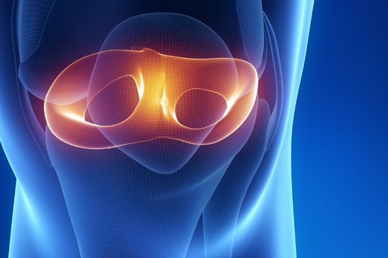
The meniscus converts axial compressive load into circumferential tensile stresses, distributed by collagen bundles oriented along the axis of the meniscal body. This function depends on the anisotropy of the tissue and on the gradient between the vascularized red zone and the avascular white zone, where healing capacity is minimal. Partial meniscectomy reduces the contact surface and accelerates chondral degeneration, while allogeneic transplants and collagen or polyurethane implants show limits in integration and durability.
An effective substitute should therefore reproduce the circumferential tensile modulus, the capacity to absorb compression, and a fibrillar organization that guides remodeling toward a functional fibrocartilaginous tissue.
The geometry of the meniscus adds a further level of complexity, because the crescent shape with a triangular cross-section and the insertion onto the tibial horns are essential to its function. An implant that does not reproduce the anatomical profile, however strong, will not distribute load correctly and risks extruding from the joint. The construction of patient-specific scaffolds, derived from magnetic resonance imaging of the contralateral meniscus and produced by three-dimensional printing or by casting in dedicated molds, is an area of active development.
Lamellar, aligned and composite scaffolds
Fibroin makes it possible to build matrices with controlled porosity and aligned fibers, using electrospinning onto a rotating collector, weaving, and infiltration into textile structures. The most convincing designs combine an oriented-fiber component, intended to bear tension, with a spongy phase that promotes cell infiltration and matrix synthesis. Gradient scaffolds have also been described, with greater porosity and angiogenic functionalization in the peripheral region and a more compact structure in the inner region, in analogy with the native vascular profile.
The comparison with native tissue must be based on compressive and tensile moduli measured under hydrated conditions, on suture retention, which is decisive for anchorage to the meniscal horns, and on resistance to prolonged cyclic loading. A matrix that holds up in a static test may fail through fatigue after thousands of cycles.
The fibrillar hierarchy of the meniscus comprises main circumferential bundles, radial tie fibers, and a superficial layer with an irregular weave. Reproducing this organization requires a layered approach, with fibroin lamellae oriented differently and subsequently assembled, so as to replicate both circumferential strength and the cohesion between bundles, which in native tissue prevents delamination and longitudinal tears. Good interlaminar cohesion is a requirement often verified with shear and peel tests.
Associated biological strategies
Seeding with mesenchymal stem cells or chondrocytes, often in combination with TGF-beta3, has shown the ability to induce a fibrochondrogenic phenotype with production of type I and type II collagen and glycosaminoglycans. Alternatively, acellular matrices functionalized with cell-recruiting factors aim to exploit synovial fluid and bone marrow as sources of progenitors. In the avascular zone, the release of angiogenic factors, or combination with techniques that provide a bleeding supply, can substantially change the outcome.
The bioreactor with mechanical stimulation, in dynamic compression and in perfusion, is a useful tool for conditioning cellularized constructs. A cyclic load of physiological magnitude stimulates matrix synthesis and fiber alignment, whereas excessive loads induce a catabolic phenotype. For constructs intended for implantation, preliminary maturation under dynamic conditions can reduce the time needed for functionalization in vivo, at the cost of greater manufacturing complexity.
Validation and next steps
Ovine and caprine models are the most appropriate for biomechanical evaluation, because load and joint dimensions approximate those of humans. Endpoints must include protection of the articular cartilage, because the true indicator of success of a meniscal implant is the prevention of osteoarthritis, not merely the survival of the implant. On this point, long-term follow-up and quantitative imaging assessments are needed.
Surgical fixation and long-term performance
In practice, one of the reasons for the failure of meniscal implants is fixation. Anchorage to the horns and peripheral suture to the capsule must withstand high cyclic loads, and the material must have adequate suture retention, with values that in vitro tests should exceed those of native tissue by a safety margin. The possibility of reinforcing the anchorage zones with denser weaving, while keeping a more compliant central portion, is a design choice consistent with function.
Long-term evaluations must verify the preservation of the implant’s geometry, the absence of extrusion, and the protection of the femoral and tibial cartilage, using measures such as chondral thickness and histology with standardized scoring. A useful observation concerns the relationship between degradation rate and remodeling: an implant that loses strength before the newly formed tissue has acquired adequate properties is exposed to failure, whereas one that persists for too long may hinder the reorganization of the tissue. The choice of optimal kinetics is not universal and must be calibrated on the animal model and, in prospect, on the patient population.
