
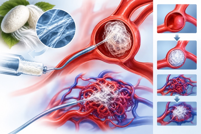
Limitations of current embolic agents
In the embolization of cerebral aneurysms, arteriovenous malformations and peripheral vascular lesions, operators use metal coils, microparticles and liquid embolics such as NBCA and ethylene-vinyl alcohol copolymers dissolved in solvent. Each has a profile that is difficult to optimize, with recanalization and coil compaction in wide-neck aneurysms, catheter adhesion and imaging artifacts for liquid embolics, and solvent toxicity in some formulations. This is where the interest in materials that gel in situ under milder conditions and integrate with the vessel wall comes from.
The choice of embolic agent depends on the lesion. In saccular cerebral aneurysms the goal is occlusion of the sac while preserving the parent vessel, and for wide necks stents or supporting balloons are used, whereas in arteriovenous malformations the agent has to penetrate the nidus all the way to the draining venous compartments, with the risk of non-target pulmonary or cerebral embolism if the material migrates. The multiplicity of scenarios makes a single solution unlikely and explains why research is turning to materials with tunable properties.
Controlled gelation and required properties
Fibroin solutions can be turned into hydrogels by sonication, pH reduction, the addition of ions or enzymatic crosslinking, with gelation times that can be tuned from seconds to hours. The advantage for endovascular use is the ability to inject a low-viscosity sol through a microcatheter and have it solidify at the target site, filling irregular geometries. The requirements to be met are gelation kinetics predictable enough to avoid catheter occlusion or distal migration, mechanical strength adequate to withstand pulsatile pressure, and radiopacity obtained with iodinated contrast agents, tantalum or bismuth nanoparticles, taking care that the additive does not alter gelation.
Rheological behavior, in particular viscosity at high shear rates and thixotropy, must be characterized under real injection conditions, with pulsatile flow in vessel models and aneurysm phantoms.
Sonication-induced gelation, which accelerates the transition toward beta sheets, is difficult to apply in the endovascular setting, and for this reason mechanisms triggered by pH changes or by the presence of calcium or potassium ions are preferred, or else those triggered by the in situ encounter with a second component. Two-component formulations, mixed at the catheter outlet or in a coaxial microcatheter, make it possible to control the onset of gelation, whereas single-component systems require far more stringent kinetics to avoid premature occlusion.
Silk textile devices
A parallel line of work concerns the use of silk thread as a thrombogenic component of coils and as a coating for flow-diverting devices. The ability to promote platelet adhesion and the formation of organized thrombus, already known in the surgical setting, is exploited to accelerate stable occlusion. The issue of surface standardization remains, in order to avoid variable inflammatory responses.
The biological response to the material is a topic of interest. The organized thrombus that forms on the device and in the aneurysm sac must evolve into stable fibrous tissue, and the persistence of loose thrombus or chronic inflammation increases the risk of recanalization. The use of bioactive materials that stimulate neointimal formation at the neck can promote definitive healing and reduce the need for prolonged antiplatelet therapy.
Evidence and development
Rabbit models, with surgically created sidewall aneurysms, are the standard for evaluating angiographic occlusion and neck healing, while malformations require swine rete mirabile models. Long-term safety, in particular the response of the vessel wall and the risk of non-target embolism, is the element on which clinical acceptability will be decided.
Safety aspects and the path to the clinic
An embolic agent comes into direct contact with blood and vessel walls, so its hemocompatibility profile, including platelet activation, complement activation and hemolysis, has to be characterized according to the ISO protocols specific to blood-contacting devices. To these tests are added the assessment of particulates, relevant because fragments released into the circulation can cause distal embolism, and behavior in magnetic resonance imaging and tomography, since the follow-up of lesions depends on image quality.
From a clinical standpoint, the first applications are likely to be in peripheral lesions and in non-cerebral therapeutic embolizations, such as venous and lymphatic malformations, where risk tolerance is broader and endpoint standardization is simpler. The experience accumulated in these settings can form the basis for a later move to interventional neuroradiology, which demands demonstrations of safety and occlusion durability at a very high level.
