
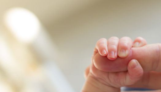
Being born before time means, among the many physiological consequences, entering the world with skin that has not yet completed its own formation. Histological analysis shows that epidermal development is completed in utero around the 34th week of gestational age. Newborns who are born before this threshold present elevated rates of transepidermal water loss (TEWL) and transcutaneous heat dispersion, with consequent difficulties in maintaining homeostasis. What for a full-term infant is an efficient protective envelope, in the premature baby is still a porous, fragile, almost transparent tissue that struggles to carry out its essential functions.
In the most immature newborns, the poorly developed epidermal barrier produces three main effects: high transepidermal water loss, increased percutaneous absorption of external substances, and greater vulnerability to mechanical trauma. These three problems intertwine in a concerning way in the neonatal intensive care environment, where the skin is continuously exposed to electrodes, adhesive patches, probes, antiseptics, and environments with variable temperature and humidity. Premature newborns who spend long periods in neonatal intensive care are particularly vulnerable to skin breakdown due to their immature epidermal barrier, and their skin is susceptible to TEWL to such a degree as to require highly effective emollients and delicate handling techniques.
The problem does not resolve quickly with simple growth. When comparing the thoracic skin of 36 premature newborns with that of 39 full-term newborns over two weeks, TEWL was found to be significantly higher in the premature infants compared to those born at term, suggesting that even at 7–8 weeks from birth, skin integrity remains compromised. This maturational delay exposes the most fragile infants to a prolonged risk of infection, fluid imbalance, and thermal injury, making urgent the search for solutions that artificially supplement what biology has not yet had the time to build.
Biocompatibility and immune response
One of the historical obstacles to the clinical use of silk proteins has been the fear of an adverse immune response. Research over recent decades has, however, considerably refined this picture. Fibroin and sericin offer attractive characteristics, including biocompatibility, tunable biodegradability, controllable tensile strength, manageability in aqueous or organic media, the ability to be functionalized and to incorporate drugs, growth factors, and other bioactive molecules, minimal inflammatory reactions in the host, wide availability, and low costs.
With regard to sericin in particular, various forms of materials such as nanoparticles, hydrogels, scaffolds, sponges, and films prepared from sericin have shown no marked immune responses or inflammatory reactions, such as mast cell degranulation. An important reason for the low immunogenicity of sericin lies in its rich content of hydrophilic amino acids. Furthermore, compared to current artificial materials such as polylactic acid or polyethylene glycol, the degradation products of sericin and fibroin are small molecular amino acids, which present a lower inflammatory response and better biocompatibility, whereas the degradation products of artificial materials such as polylactic acid produce a marked inflammatory response by lowering the pH of the environment.
Purified fibroin, obtained after the removal of sericin through the degumming process, has been recognized by the US FDA as an approved biomaterial for medical use. Silk fibroin is an FDA-approved biomaterial of emerging importance in the fields of regenerative science and tissue engineering. This regulatory recognition is relevant because it maps out a clinical transfer pathway for pediatric and neonatal applications.
Cell proliferation, healing, and antimicrobial defense
Silk proteins do not simply serve as an inert physical barrier, but actively interact with the cells of the host tissue, modulating their behavior in directions favorable to regeneration. Sericin has been shown to stimulate cell adhesion and proliferation, facilitating cell growth, as well as cell differentiation toward specific cell types.
From a molecular standpoint, fibroin stimulates the expression of genes in the NF-κB signaling pathway, leading to increased expression of vimentin, fibronectin, cyclin D1, and VEGF. SF also stimulates the MEK, PI3K, and JNK pathways, resulting in increased cell migration. These mechanisms are fundamental in tissue repair processes: VEGF promotes neovascularization, fibronectin promotes cell adhesion and keratinocyte migration, and cyclin D1 regulates the cell cycle, accelerating epidermal regeneration.
On the antimicrobial front, although fibroin alone does not have significant intrinsic antibacterial activity, composite materials combining it with other agents show promising results. Sericin possesses antioxidant and antimicrobial activity. Silk films produced from the whole cocoon in CaCl?-ethanol-H?O solution showed significant inhibition against Escherichia coli (23.1 mm) and Staphylococcus aureus (20.2 mm). For premature newborns, who are exposed to hospital pathogens such as Staphylococcus and Klebsiella in a NICU setting, this property has direct clinical value. A carboxymethylcellulose/sericin-based hydrogel was found to be capable of reducing the expression of IL-1β, IL-6, and TNF-α, improving the pro-inflammatory response at the wound site. Modulating the inflammatory response is particularly important in premature neonatal skin, where an excessive or poorly regulated inflammatory response can worsen the tissue damage already present.
The biomedical forms of silk proteins
One of the reasons that make silk proteins so well suited to being transformed into artificial skin barriers is their extraordinary processing versatility. Fibroin can be used alone or in combination with other materials in various scaffold forms, such as nanofiber membranes, hydrogels, sponges, or films, adaptable to specific applications.
Thin films of fibroin — or fibroin/sericin blends — are among the most studied formulations for superficial cutaneous applications. Restructured SF/SS films have proven to be stable, transparent, endowed with good mechanical properties, antibacterial activity, and cytocompatibility, and can therefore serve as suitable biomaterial candidates for skin regeneration applications. Transparency is a non-trivial feature in the neonatal context: allowing direct visualization of the underlying skin without removing the dressing is essential for the continuous monitoring that characterizes neonatal intensive care. Fibroin-based hydrogels deserve particular mention for their ability to maintain a moist microenvironment and to release bioactive molecules in a controlled manner. Fibroin and sericin offer the ability to be functionalized and to incorporate drugs, growth factors, and other bioactive molecules. In a neonatal context, this property could allow the incorporation of epidermal growth factors or antimicrobial agents for local release, reducing the need for systemic treatments with their associated side effects.
