
Fibroin-based bioprinting has long since ceased to be a laboratory curiosity and has established itself as one of the most versatile platforms for the fabrication of three-dimensional tissue constructs. The reason for this success does not lie in a single outstanding property, but rather in the combination of aqueous processability, mechanical tunability across several orders of magnitude, and highly controllable gelation kinetics. Researchers working on the reconstruction of complex cellular microenvironments seek precisely these characteristics: a material that enables the decoupling of print fidelity from cell viability while simultaneously allowing the engineering of stiffness gradients, vascular geometries, and biochemical niches within the same construct. These aspects, rather than the general properties of the biopolymer itself, deserve particular attention.
Rheological behavior and the biofabrication window
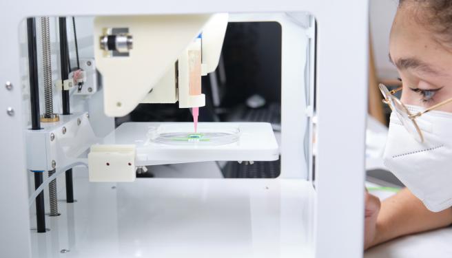
Extrusion-based printing, still the most widespread technique for silk fibroin bioinks, depends on a delicate balance. The ink must behave as a yield-stress fluid, flowing readily under the shear stress imposed within the nozzle and then rapidly recovering its structure after deposition to support subsequent layers without collapse. Pure aqueous fibroin solutions are generally insufficiently viscous to guarantee such shape fidelity, which is why a wide range of functionalization and composite formulation strategies has emerged. Modulating protein concentration, controlling molecular weight distributions through degumming time, or incorporating viscosity-enhancing agents allows researchers to move within what is commonly referred to in the literature as the biofabrication window: the range of parameters where a construct is simultaneously printable and biologically permissive.
The critical challenge is that conditions improving printability—high concentrations, elevated shear stresses, and rapid gelation—are often the same conditions that place encapsulated cells under stress. The conformational transition from random coil to β-sheet structures, which imparts water stability to fibroin, can be triggered by mechanical stress and local dehydration during extrusion, potentially leading to uncontrolled gelation at the nozzle tip. Designing a high-performance fibroin bioink therefore requires precise control of crystallization kinetics, slowing them when deposition fidelity is needed and accelerating them when post-print stabilization becomes necessary.
Physical crosslinking mechanisms and control of β-Sheet content
Physical crosslinking remains the most cytologically conservative strategy because it does not introduce reactive species or potentially toxic reagents. The formation of crystalline Silk II domains can be induced through sonication, high-speed stirring, pH reduction, exposure to alcohols, or treatment with lyotropic salts. Each of these stimuli shifts the conformational equilibrium toward intermolecular β-sheet aggregation, which acts as the physical crosslinking nodes of the network and imparts stiffness, water insolubility, and resistance to enzymatic degradation.
For the construction of cellular microenvironments, the β-sheet content becomes an almost continuously tunable design parameter. By regulating the degree of crystallization, it is possible to simultaneously control elastic modulus, swelling ratio, and proteolytic degradation rate, enabling the assembly of constructs containing stiff, minimally swelling regions adjacent to softer, more hydrated domains. Recent strategies have exploited this relationship to fabricate self-folding bilayer constructs in which a sonicated, highly crystalline layer serves as a passive substrate for a more compliant active layer. The historical drawback of physical crosslinking—namely, slow gelation kinetics that are difficult to synchronize with deposition—has been mitigated through additives such as glycerol, which induces thermosensitive behavior and accelerates the sol-gel transition without requiring chemical crosslinkers.
Enzymatic crosslinking and dityrosine bond formation
When the goal is to obtain an elastic, optically transparent hydrogel formed under strictly physiological conditions, enzymatic crosslinking mediated by horseradish peroxidase (HRP) in the presence of hydrogen peroxide has become a benchmark approach. The enzyme catalyzes the oxidative coupling of tyrosine residues along the protein chain, generating covalent dityrosine bonds that crosslink the network within seconds to minutes, depending on enzyme and peroxide concentrations. The resulting material is highly extensible and markedly different from the brittle, β-sheet-rich gels obtained through physical crosslinking, making it particularly suitable for soft tissues subjected to cyclic deformation.
Its relevance to three-dimensional cellular microenvironments is twofold. First, rapid gelation stabilizes the printed geometry before gravitational relaxation or diffusive spreading can occur. Second, because crosslinking proceeds under physiological temperature and pH conditions, cells and growth factors can be directly encapsulated within the precursor solution, yielding densely cellularized constructs with homogeneous distributions. Careful management of hydrogen peroxide concentration remains essential, however, since excessive levels can induce oxidative stress. Furthermore, tyrosine residues themselves can act as free-radical scavengers, a factor that becomes particularly significant when enzymatic chemistry is combined with photopolymerization processes.
Methacrylated fibroin and light-based printing
The most significant advance in printing resolution has been achieved through methacrylation of the fibroin protein chain. By reacting fibroin with glycidyl methacrylate, methacrylate groups are introduced onto lysine amino groups and hydroxyl functionalities, producing a photocrosslinkable derivative commonly known as SilMA (Silk Methacrylate). In the presence of a photoinitiator such as lithium phenyl-2,4,6-trimethylbenzoylphosphinate (LAP) and exposure to near-ultraviolet or violet light—typically at wavelengths around 365 or 405 nm—the methacrylate groups undergo radical polymerization to form a stable covalent network. The landmark study by Kim and colleagues in 2018 demonstrated that such modified fibroin bioinks could be printed with high precision and biocompatibility using digital light processing (DLP) technology.
Light-based printing, whether implemented through DLP or stereolithography, fundamentally changes the fabrication paradigm compared with extrusion. Rather than depositing material filament by filament, a volume of precursor is selectively polymerized layer-by-layer—or even volumetrically—achieving resolutions that are difficult to attain mechanically and removing constraints imposed by nozzle diameter. This enables the fabrication of microchannels, interconnected lattices, and precisely controlled porous architectures, all of which are essential for reproducing the spatial complexity of native microenvironments.
An important and somewhat counterintuitive aspect must be considered: fibroin's ability to scavenge free radicals, arising from its tyrosine residues, interferes with radical photopolymerization kinetics. Rather than being merely a limitation, this effect can be exploited to restrict radical diffusion beyond the irradiated region, thereby improving edge sharpness and enhancing the effective resolution of printed constructs.
Composite bioinks, interpenetrating networks, and double-network hydrogels
Silk fibroin is rarely used alone when engineering complex cellular microenvironments. The established practice is to formulate composite systems that compensate for its limitations, particularly its relatively modest cell adhesiveness and its intrinsically limited printability. Pairing fibroin with gelatin—or its methacrylated derivative, GelMA—is perhaps the most common strategy. Gelatin contributes cell-adhesion motifs and thermoreversible behavior advantageous for deposition, while fibroin provides long-term structural stability and mechanical tunability.
Combinations with methacrylated hyaluronic acid, alginate, methacrylated alginate, or carrageenan further expand the accessible property space, each introducing an orthogonal crosslinking chemistry that complements that of fibroin.
From an engineering perspective, the most intriguing opportunity lies in the creation of interpenetrating polymer networks (IPNs) and double-network architectures, where two crosslinking mechanisms coexist within the same volume. For example, radical photopolymerization can be followed by sonication-induced β-sheet formation, producing materials that combine the exceptional mechanical robustness characteristic of double networks with the biocompatibility of processes fully compatible with cell survival. This modular design philosophy is precisely what makes fibroin particularly suitable for fabricating heterogeneous niches: by locally varying composition, crosslinking chemistry, and crystallization degree, regions with distinct mechanical and biochemical identities can be sculpted within a single construct.
Microenvironment engineering: gradients, stiffness, and mechanotransduction
The promise of fibroin bioprinting extends far beyond simply depositing cells in three dimensions; it lies in controlling the physical and chemical context that governs cellular fate. Matrix stiffness is among the most powerful signals perceived by cells. Variations in elastic modulus direct stem cell differentiation, regulate intracellular mechanotransduction pathways, and influence cell migration and tissue organization.
Because fibroin stiffness depends directly on β-sheet content and crosslinking density—and because both parameters can be spatially modulated—it becomes possible to print stiffness gradients that recapitulate native tissue interfaces, such as the osteochondral junction between cartilage and bone.
Mechanical control is complemented by architectural control. The ability to fabricate interconnected channel networks, particularly accessible through light-based technologies, addresses one of the longstanding bottlenecks in tissue engineering: perfusion and prevascularization of thick constructs. Without an efficient mass transport network, cells in internal regions suffer from oxygen and nutrient deprivation long before functional vascularization can develop. Printing microchannels within fibroin matrices, potentially lining them with endothelial cells through co-culture approaches, represents one of the most promising strategies for overcoming the thickness limitations that continue to separate many engineered constructs from physiological relevance. Multimaterial printing provides an additional degree of freedom by allowing cellularized deposits to be combined with sacrificial or support materials that are subsequently removed to create patent luminal structures.
Cell Viability, processing stress, and degradation
Every formulation and processing decision ultimately affects the health of encapsulated cells, and this is where fibroin exhibits one of its strongest advantages. Most fibroin crosslinking chemistries operate in aqueous environments under mild conditions, minimizing exposure to organic solvents and harmful temperatures.
Nevertheless, two recurring challenges remain. The first is shear stress during extrusion, which increases with viscosity and decreasing nozzle diameter and can compromise cell membranes precisely when higher printing resolution is sought. The second is the chemical stress associated with radical-mediated crosslinking, where photoinitiators and ultraviolet exposure generate reactive oxygen species with potential cytotoxic effects. The use of highly efficient visible-light photoinitiators at low concentrations, together with the radical-scavenging activity of tyrosine residues, helps mitigate these risks.
Finally, degradation must be designed rather than tolerated. Silk fibroin undergoes proteolytic degradation, and its degradation rate depends predictably on β-sheet content: highly crystalline matrices persist longer, whereas more amorphous structures are remodeled rapidly. Synchronizing degradation kinetics with the deposition of newly synthesized extracellular matrix by resident cells is what distinguishes a simple scaffold from a truly regenerative construct. Ideally, the printed material gradually relinquishes both space and mechanical function to the tissue being formed by the cells themselves, thereby avoiding both premature resorption and prolonged persistence as a foreign body.
