
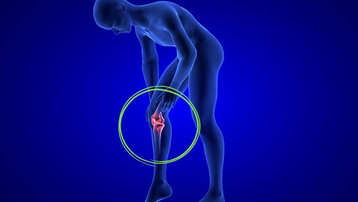
Articular cartilage represents one of the most complex tissues to regenerate in the human organism, characterized by a limited intrinsic capacity for self-repair due to its avascular nature and absence of innervation. Osteoarthritis, a degenerative pathology affecting millions of people worldwide, finds its most devastating manifestation precisely in the progressive erosion of this tissue. In this context, hybrid hydrogels composed of silk fibroin and collagen emerge as extraordinarily promising biomaterial platforms, capable of replicating with increasing fidelity the architecture and biomechanical properties of native cartilage.
The scientific rationale of the silk-collagen combination
The synergy between silk fibroin and collagen is not coincidental, but responds to precise biomechanical and biological necessities. Fibroin, a structural protein extracted primarily from the silkworm Bombyx mori, is distinguished by exceptional mechanical properties deriving from its crystalline structure organized in antiparallel beta-sheets. This molecular conformation confers to the material a tensile strength comparable to that of steel on a weight basis, a fundamental characteristic for withstanding the mechanical loads typical of joints. Collagen, particularly type II predominant in articular cartilage, instead provides the essential biological signals for cellular recognition and adhesion of chondrocytes, the specialized cells in the production of cartilaginous matrix.
When these two components are integrated into a three-dimensional hydrogel matrix, a biomaterial is obtained that combines the mechanical robustness of silk with the intrinsic bioactivity of collagen. Mechanical characterization studies have demonstrated that hybrid hydrogels can achieve elastic moduli ranging between 0.1 and 1 MPa, an interval that approaches significantly the values of human articular cartilage, typically in the order of 0.5-0.9 MPa in the superficial zones. This mechanical correspondence is crucial because substrate stiffness profoundly influences the differentiative fate of mesenchymal stem cells, orienting them toward the chondrocytic phenotype when the microenvironment presents appropriate elastic properties.
Porous architecture and biomimetic design of scaffolds
The design of three-dimensional scaffolds for cartilage regeneration requires particular attention to porosity and interconnectivity of the structure. Silk-collagen hydrogels can be fabricated through various methodologies, including controlled lyophilization, electrospinning, and more advanced three-dimensional bioprinting techniques. Lyophilization, in particular, allows the creation of architectures with porosity exceeding 90% and pore dimensions variable between 50 and 300 micrometers, optimal parameters for allowing cellular migration, nutrient diffusion, and elimination of metabolites.
The interconnectivity of pores represents an often underestimated but essential aspect: it is not enough for the scaffold to be porous, it is necessary that the pores be connected to each other to create true biological highways through which cells can move and colonize the entire structure. In optimized hybrid hydrogels, this interconnectivity reaches values exceeding 95%, guaranteeing homogeneous cellular distribution and uniform extracellular matrix production throughout the scaffold volume. The morphology of pores can be further modulated by varying the ratio between fibroin and collagen: compositions with higher fibroin content tend to generate more elongated and oriented pores, mimicking the structural anisotropy of native cartilage, where collagen fibers are organized in layers with specific orientation from the deep surface to the calcified zone.
Mechanical properties and viscoelastic behavior
Articular cartilage is not simply a rigid tissue, but a sophisticated material with viscoelastic behavior, capable of dissipating energy under cyclic load and gradually recovering its original form after deformation. This property is fundamental for absorbing mechanical shocks during locomotion and preventing damage to the underlying bone surfaces. Silk-collagen hybrid hydrogels have demonstrated the ability to replicate this complex viscoelastic behavior, exhibiting an elastic component due mainly to the beta crystals of fibroin and a viscous component deriving from the intermolecular interactions of collagen and the aqueous content of the matrix.
Cyclic compression tests have revealed that these biomaterials can withstand up to one million load cycles without significant structural degradation, an indispensable requirement considering that a knee joint, for example, can be subjected to several million cycles during normal lifespan. The resistance to mechanical fatigue can be further enhanced through controlled crosslinking treatments, using agents such as genipin or EDC (1-ethyl-3-(3-dimethylaminopropyl)carbodiimide), which create covalent bonds between protein chains without introducing cellular toxicity. The degree of crosslinking must be carefully balanced: excessive crosslinking stiffens the matrix too much, compromising cellular migration, while insufficient crosslinking can lead to premature collapse of the scaffold under physiological load.
Cellular interactions and chondrogenic microenvironment
The success of a scaffold for cartilage regeneration depends critically on its capacity to support survival, proliferation and especially the maintenance of the chondrocytic phenotype of cells. Chondrocytes represent in fact an extremely sensitive cellular population to the surrounding microenvironment, with a tendency toward dedifferentiation when cultured in inadequate conditions, losing the capacity to produce type II collagen and cartilage-specific proteoglycans to instead synthesize type I collagen typical of fibrous tissue.
Silk-collagen hydrogels have demonstrated exceptional capabilities in preserving the chondrocytic phenotype, as evidenced by sustained expression of specific markers such as Sox9, a key transcription factor in chondrogenesis, and aggrecan, the principal proteoglycan of cartilaginous matrix. This superior performance is attributable to multiple synergistic factors: the presence of collagen provides specific adhesion sites through RGD sequences (arginine-glycine-aspartic acid) that are recognized by membrane integrins, while the three-dimensional structure of the hydrogel recreates that condition of "cellular compression" typical of native cartilage, where chondrocytes reside in lacunae surrounded by dense matrix. This geometry favors cell-cell and cell-matrix interactions that are fundamental for the maintenance of the differentiated phenotype.
Swelling properties and nutrient permeability
An aspect often neglected in the design of cartilaginous scaffolds concerns swelling properties and the capacity to retain water. Articular cartilage contains approximately 70-80% water by weight, and this hydric component is essential for the lubricating properties and for the compression resistance of the tissue. Silk-collagen hydrogels can absorb significant quantities of fluid, with swelling degrees variable between 500% and 1500% depending on composition and degree of crosslinking. This swelling behavior must be optimized to avoid two problematic extremes: excessive swelling can lead to a critical dilution of the matrix with loss of mechanical properties, while insufficient swelling limits the diffusion of nutrients and elimination of metabolic waste.
The permeability of the scaffold represents a complementary parameter to swelling and determines the velocity with which molecules such as glucose, oxygen and growth factors can diffuse through the matrix to reach incorporated cells. In optimized hybrid hydrogels, the diffusion coefficient for small molecules like glucose approaches that measured in native cartilage, guaranteeing adequate nutritional supply even in the central regions of constructs of clinically relevant dimensions, typically in the order of several millimeters of thickness. This characteristic is particularly important considering the avascularity of cartilage, where all metabolic exchanges must occur by diffusion through the matrix from the surrounding synovial fluid.
Incorporation of bioactive factors and controlled release
The functionalization of silk-collagen hydrogels with bioactive molecules represents an advanced strategy to enhance their regenerative potential. TGF-β3 (transforming growth factor beta-3) is recognized as one of the most potent inducers of chondrogenesis, stimulating the synthesis of proteoglycans and type II collagen. The incorporation of this growth factor into the hydrogel matrix, however, presents significant challenges related to the preservation of its bioactivity and control of release kinetics. Silk fibroin offers unique advantages in this context, thanks to the capacity to stabilize proteins through electrostatic interactions and hydrogen bonds, protecting them from denaturation and enzymatic degradation.
Sophisticated release systems have demonstrated the possibility of obtaining biphasic release profiles, with an initial burst release in the first 48-72 hours that provides immediate chondrogenic stimulation, followed by sustained release for several weeks that maintains a pro-chondrogenic microenvironment during the critical phase of scaffold colonization and matrix deposition. Beyond TGF-β3, other bioactive factors of interest include BMP-7 (bone morphogenetic protein-7), which has demonstrated protective effects on arthritic chondrocytes by reducing the expression of degradative metalloproteases, and IGF-1 (insulin-like growth factor-1), which stimulates proteoglycan synthesis and inhibits chondrocyte apoptosis.
Approaches with mesenchymal stem cells
While the use of autologous chondrocytes represents the most intuitive cellular approach for cartilage regeneration, this strategy presents significant practical limitations related to the scarce availability of harvestable healthy cartilaginous tissue, donor site morbidity and the limited proliferative capacity of chondrocytes in culture. Mesenchymal stem cells (MSC), derivable from easily accessible sources such as bone marrow, adipose tissue or cord blood, offer a promising alternative thanks to their capacity to expand in vitro while maintaining differentiative potential and their capacity to differentiate toward the chondrocytic lineage in response to appropriate stimuli.
Silk-collagen hydrogels have demonstrated providing a particularly favorable microenvironment for MSC chondrogenesis, superior to that of many other tested biomaterials. Comparative studies have revealed that MSC seeded in these scaffolds express higher levels of Sox9 and collagen II compared to cells cultured in three-dimensional pellets, the in vitro gold standard for chondrogenesis, when exposed to a chondrogenic cocktail containing TGF-β3, dexamethasone and ascorbic acid. The percentage of cells that successfully complete chondrogenic differentiation can exceed 70-80% in optimized conditions, with a production of glycosaminoglycans quantitatively comparable to that of mature chondrocytes. A critical aspect concerns the prevention of terminal chondrocyte hypertrophy, a process that normally precedes endochondral ossification during skeletal development but that must be avoided in the regeneration of permanent articular cartilage. The optimal composition of the hydrogel, with carefully calibrated silk-collagen ratios, seems to contribute to maintaining cells in a stable chondrocytic state without progression toward hypertrophic phenotype.
Biodegradability and matrix remodeling
A fundamental requirement for any tissue scaffold is that its degradation be synchronized with the synthesis of new extracellular matrix by cells, so that the mechanical properties of the construct are maintained during the entire regenerative process. Silk-collagen hydrogels present modulable degradation kinetics through various molecular design strategies. Collagen is naturally susceptible to enzymatic degradation by matrix metalloproteases (MMP) and collagenases produced by chondrocytes themselves, with in vivo degradation times typically in the order of 4-8 weeks depending on the degree of crosslinking. Silk fibroin, on the contrary, presents greater resistance to enzymatic degradation, with in vivo residence times that can extend for several months.
This difference in degradative kinetics does not necessarily represent a disadvantage, but can be strategically exploited to create scaffolds with graded degradation: collagen provides immediate biological support and is progressively replaced by neosynthesized matrix, while the silk component maintains the mechanical integrity of the scaffold in the more advanced phases of regeneration when the new matrix is still maturing and acquiring definitive mechanical properties. Studies in animal models have documented how the residual percentage of scaffold at 12 weeks from implantation can vary from 20% to 60% depending on the initial silk-collagen ratio, with an inverse correlation between silk content and degradation velocity. The degradation of the scaffold releases bioactive peptides deriving from both silk and collagen, some of which have demonstrated possessing chemotactic activity for endogenous progenitor cells present in the synovial tissue, thus contributing to recruiting autologous regenerative resources.
Experimental models of osteoarthritis and preclinical results
The validation of the efficacy of silk-collagen hydrogels in the context of osteoarthritis requires experimental models that faithfully recreate the pathological characteristics of human disease. The most commonly used model is the surgical induction of osteoarthritis through destabilization of the medial meniscus in rodents or lagomorphs, which produces progressive degenerative changes in articular cartilage over the course of 8-12 weeks. Studies that have applied silk-collagen hydrogel implants in chondral defects created in this context have documented encouraging results in terms of defect filling, integration with surrounding tissue and quality of repaired cartilage.
Histological analyses at 12-24 weeks post-implantation have revealed the formation of reparative tissue with histomorphological characteristics similar to native hyaline cartilage, including the presence of chondrocytes organized in columns in the deep zones and the accumulation of matrix rich in proteoglycans demonstrable with specific stains such as Safranin-O. Histological scores according to internationally validated scales, such as the ICRS (International Cartilage Repair Society) score, reach values significantly superior compared to untreated defects or those treated with control materials, with some studies reporting scores in the interval 10-12 out of 14, indicative of almost complete regeneration. Immunohistochemical analyses confirm the prevalent expression of type II collagen rather than type I, a crucial indicator of the hyaline rather than fibrous nature of the repaired tissue, which determines the long-term functional durability of the repair.
Tribological properties and articular lubrication
An aspect of cartilaginous functionality often neglected in biomaterials research is represented by tribological properties, that is the capacity to provide surfaces with low friction coefficient that permit articular sliding without wear. Healthy articular cartilage presents one of the lowest friction coefficients known in nature, typically in the interval 0.001-0.03 when lubricated by synovial fluid, surpassing the performance of many engineered materials. This exceptional lubrication derives from the combination of various mechanisms including boundary lubrication provided by lubricin (PRG4), a glycoprotein present in the cartilaginous surface, and hydrodynamic lubrication mediated by the pressurization of interstitial fluid during loading.
Mature silk-collagen hydrogels, after cellular colonization and matrix deposition, have demonstrated developing tribological properties that approach those of native tissue. Tribometric tests conducted in the presence of synovial simulant reveal friction coefficients in the interval 0.05-0.15 for constructs of 8-12 weeks, values that, although still superior to native cartilage, represent a substantial improvement compared to fibrous reparative tissues that typically present coefficients exceeding 0.3. The progressive improvement of tribological properties correlates with the superficial accumulation of highly hydrated proteoglycans and with the initiation of lubricin production by differentiated chondrocytes, suggesting that even more mature constructs could reach tribological performance indistinguishable from the original tissue.
Bioprinting strategies for patient-specific geometries
The advent of three-dimensional bioprinting technologies has opened revolutionary possibilities in the fabrication of cartilaginous scaffolds with complex and personalized geometries based on patient diagnostic imaging. Silk-collagen hydrogels can be formulated as bio-ink with appropriate rheological properties for extrusion through nozzles of variable dimensions, typically in the interval 200-400 micrometers, permitting layer-by-layer construction of three-dimensional structures with submillimetric resolution. The viscosity of the bio-ink must be carefully balanced: it must be sufficiently low to permit extrusion without generating excessive pressures that would damage potentially incorporated cells, but sufficiently high to maintain the printed form before complete gelification.
One of the most promising applications of this technology concerns the creation of stratified osteochondral implants that recreate the entire functional unit of cartilage-subchondral bone. By varying the composition of the bio-ink between layers, it is possible to generate constructs with a continuous gradient of properties, passing from a composition rich in collagen II in the superficial regions to a composition enriched with calcium phosphate and collagen I in the deep regions destined for integration with subchondral bone. Pilot studies have demonstrated the feasibility of printing complex geometries such as femoral condyles or tibial plateaus with dimensional fidelity superior to 95% compared to the original digital model, opening concrete prospects for on-demand fabrication of personalized implants for extensive cartilaginous defects where conventional therapeutic options are limited.
Immunogenicity and inflammatory response
The biocompatibility of any implantable biomaterial is not limited to the sole absence of direct cytotoxicity, but also includes the evaluation of the immune and inflammatory response that the material can trigger. This aspect is particularly relevant in the articular context, where an excessive or prolonged inflammatory response can exacerbate degenerative processes and compromise tissue regeneration. Silk fibroin, in its native form, contains an adhesive protein called sericin that can provoke immunogenic reactions; therefore, preparation protocols include degumming steps to remove this component. Degummed fibroin has demonstrated excellent biocompatibility with minimal inflammatory responses after implantation.
Immunogenicity studies have evaluated the cellular response to silk-collagen hydrogels through analysis of immune cell infiltration, production of pro-inflammatory cytokines such as IL-1β, IL-6 and TNF-α, and formation of fibrous encapsulation. The results indicate that these biomaterials induce a transitory and self-limiting foreign body reaction in the first 2-4 weeks post-implantation, characterized by infiltration of macrophages and neutrophils, followed by rapid resolution of inflammation and progressive integration with host tissue. The expression of pro-inflammatory cytokines reaches a peak at 7-10 days post-implantation and then declines to basal levels within 4 weeks, a temporal profile compatible with a normal healing response rather than with a chronic pathological reaction. The absence of dense fibrous capsule formation, which would represent a barrier to tissue integration, is a positive indicator of the superior biocompatibility of these hybrid materials.
