
Chronic perforation of the tympanic membrane is one of the most common conditions in otologic practice, with consequences ranging from conductive hearing loss to recurrent otorrhea and a progressive deterioration in the patient's quality of life. The surgical reconstruction of this structure, historically entrusted to autologous grafts of temporalis fascia or perichondrium, has represented the reference standard for decades, delivering high success rates but requiring the surgeon to harvest tissue from a donor site and imposing an additional operative burden on the patient. It is within this clinical space, where the demand for ever less invasive solutions meets the limitations of traditional materials, that silk fibroin proposes itself as a synthetic biomaterial capable of redefining the approach to reconstructive middle ear surgery.
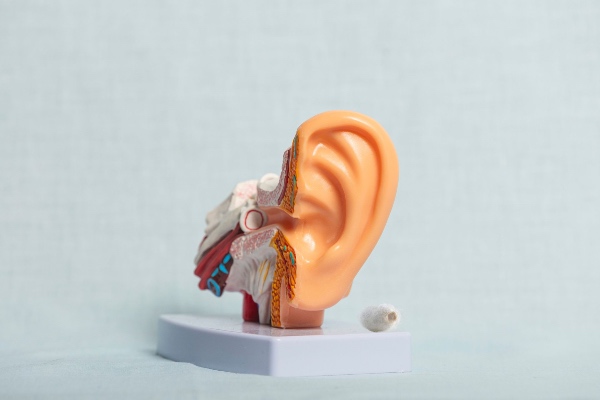
The tympanic membrane is an anatomical structure of extraordinary mechanical refinement. Its thickness, on the order of a few tens of microns, conceals a trilaminar organization in which an outer epidermal layer, an intermediate fibrous layer, and an inner mucosal layer cooperate to transform acoustic pressure waves into mechanical vibrations transmitted to the ossicular chain. The fibrous layer, in particular, owes its function to an ordered network of collagen fibers arranged in radial and circumferential directions, an architecture that grants the membrane the stiffness and elasticity needed to respond linearly across a broad spectrum of frequencies. Any material intended to replace or regenerate this structure must confront this twofold requirement, providing adequate mechanical support without dampening the vibratory capacity that underlies auditory function.
The properties of fibroin relevant to the middle ear
Fibroin, a structural protein extracted from silkworm cocoons and purified through degumming processes that remove the sericin component, presents a property profile that makes it particularly well suited to this context. Its secondary structure, dominated by crystalline beta-sheet domains interspersed with amorphous regions, determines a mechanical behavior that combines tensile strength and flexibility in a balance rarely found among conventional synthetic polymers. From this protein it is possible to obtain thin, transparent, and optically homogeneous films, whose thickness can be precisely modulated on the order of microns through substrate casting or controlled deposition techniques.
Transparency represents an operative advantage that is anything but secondary. A transparent graft allows the surgeon to visualize the correct apposition of the margins over the perforation and to verify the absence of collections or overlaps during placement, an element of control that opaque autologous tissues do not offer. Accompanying this visual quality is a biocompatibility documented by an extensive body of literature on fibroin employed across disparate fields of regenerative medicine, where the protein proves capable of supporting cell adhesion and proliferation without triggering significant inflammatory responses or clinically meaningful foreign body reactions.
The tunability of the degradation rate constitutes a further point of interest. By acting on the material's degree of crystallinity, achievable through physical treatments such as exposure to water vapor or immersion in alcoholic solutions that promote the transition toward the beta-sheet conformation, it is possible to engineer films that remain in place for weeks or for months. This capacity to tune the resorption time window makes it possible to conceive of the graft not as a permanent prosthesis but as a transient scaffold, meant to guide the regeneration of the native tissue before yielding to the newly formed membrane.
Fibroin as a scaffold for tympanic regeneration
The paradigm that makes fibroin interesting in myringoplasty is not one of replacement, but rather one of regenerative guidance. A fibroin film positioned to cover the perforation functions as a biological bridge across which the epithelial cells at the margins can migrate, progressively colonizing the surface of the material and reconstituting the continuity of the membrane. The microarchitecture of the film, which can be engineered to present controlled porosity or oriented surface patterns, offers the cells a topographic substrate that directs their migration and favors their organization according to geometries that echo the native orientation of the fibers.
Numerous research groups have explored the possibility of enriching these scaffolds with growth factors, in particular with molecules belonging to the epidermal growth factor and fibroblast growth factor families, which accelerate epithelial proliferation and the deposition of connective matrix. Fibroin lends itself to this enrichment because its structure allows for the incorporation and controlled release of bioactive agents, transforming the simple mechanical support into a device capable of actively orchestrating the repair process. From this perspective, fibroin myringoplasty sits at the intersection of surgery and tissue engineering, where the operative gesture is limited to establishing the conditions that allow the tissue itself to complete the reconstruction.
The advantage of minimal invasiveness
The minimally invasive dimension is perhaps the trait that best defines the clinical potential of this approach. The availability of a standardized synthetic material eliminates at the root the need for autologous harvesting, with the consequent reduction in operative times, in the overall invasiveness of the procedure, and in the risk of donor site morbidity. An industrially produced fibroin film arrives in the operating room ready for use, with reproducible geometric and mechanical characteristics, freeing the outcome from the intrinsic variability of harvested tissue and from the learning curve that the harvesting itself entails.
This standardization also opens the way to ambulatory procedures performed via a transcanal route, without the retroauricular or endaural incisions required for access to the harvest site. In a context where otology is moving toward increasingly conservative interventions, often conducted under endoscopic control through the external auditory canal, the compatibility of a thin, flexible, and easily handled graft with these techniques represents an important element of synergy. The surgeon can introduce and position the film through small-caliber instruments, reducing tissue trauma and favoring a more rapid postoperative recovery.
