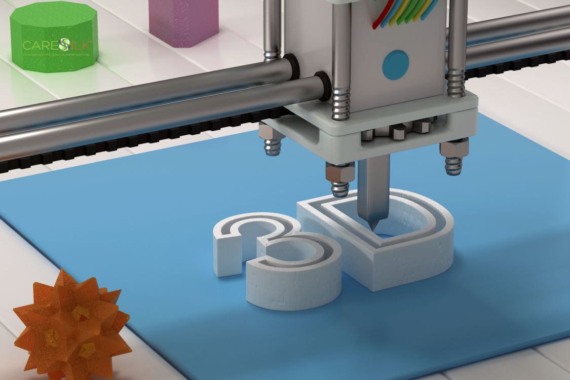
In recent years, academic and industrial research has made giant strides in using natural biomaterials for increasingly sophisticated and promising applications. Fibroin, the main structural protein of silk produced by the Bombyx mori silkworm, possesses characteristics that make it exceptionally suitable as a bioink for three-dimensional bioprinting, thanks to its extraordinary combination of biocompatibility, mechanical strength, and ability to be manipulated into various physical forms. The true revolution lies in its chemical and structural versatility. A team of researchers at Tufts University Silklab, led by Professor David Kaplan, has demonstrated how fibroin can be processed into hydrogels with controllable rheological precision, allowing optimization of viscosity and flow properties necessary for extrusion through micrometric nozzles. This fine control of physical properties is fundamental for maintaining structural integrity during the printing process, a critical problem that has limited the use of many other natural biopolymers. Studies published in Nature Materials have highlighted how the secondary structure of fibroin can be manipulated through pH control, temperature, and ionic concentration, allowing controlled transitions from random coil conformation to beta-sheet structure, responsible for the solidification of the material after deposition.
When silk promotes cell growth
Silk's compatibility with living cells perhaps represents its most significant advantage in the context of regenerative medicine. Research conducted at the Massachusetts Institute of Technology and Harvard Medical School has demonstrated that fibroin scaffolds can effectively support the adhesion, proliferation, and differentiation of multiple cell types, including fibroblasts, keratinocytes, mesenchymal stem cells, and chondrocytes. This cellular versatility is crucial when considering multi-material and multi-cellular bioprinting approaches for creating complex tissues. Particularly promising are the results obtained in 2023 by the research group of Professor Jinke Zhang at Zhejiang University, which developed a fibroin-based bioink containing hydroxyapatite nanoparticles for printing bone scaffolds with excellent osteoconductive properties, demonstrating significant mineralization and formation of new bone tissue in murine models after just eight weeks of implantation.
Controlled degradability and regenerative medicine
The controlled degradability of silk proteins represents another fundamental aspect for applications in regenerative medicine. Unlike many synthetic polymers, fibroin undergoes enzymatic degradation that can be modulated through chemical or structural modifications of the protein. Studies conducted at Imperial College London have precisely mapped the degradation profiles of silk scaffolds with different percentages of crystalline structure, demonstrating how it is possible to program degradation times from a few weeks to over a year. This characteristic is essential for synchronizing the reabsorption of the biomaterial with tissue regeneration processes, a delicate balance that largely determines the success of the implant.
The most recent developments in the field concern the chemical functionalization of fibroin to incorporate bioactive molecules that guide specific cellular behaviors. Researchers at ETH Zurich have developed conjugation methods that allow growth factors such as BMP-2, VEGF, and TGF-β to be covalently bound to the protein structure, thus creating bioactive scaffolds that not only provide structural support but also localized biochemical signals. Results published in Science Advances in October 2023 demonstrated how these 3D-printed bioactive scaffolds can induce specific and localized cellular responses, paving the way for zonally functionalized tissue constructs for the regeneration of complex tissue interfaces such as osteochondral.
Multi-nozzle systems and hybrid approaches
The research group directed by Professor Jennifer Lewis at Harvard has developed multi-nozzle bioprinting systems that allow the simultaneous deposition of different bioinks with micrometric precision. These technological advancements enable the creation of complex structures that replicate the heterogeneous architecture of native tissues. Particularly interesting is the integration of fibroin with other biomaterials such as collagen, alginate, or synthetic hydrogels in hybrid printing approaches that combine the superior mechanical properties of silk with the biochemical characteristics of other materials, thus optimizing both the printability and biological functionality of the final construct.
Clinical applications of 3D-printed fibroin-based structures are also beginning to emerge, although most remain in the preclinical experimentation phase. Particularly promising results have been obtained in the regeneration of articular cartilage, where fibroin scaffolds loaded with chondrocytes have demonstrated excellent integration and tissue regeneration in porcine models. In the field of skin regeneration, 3D-printed constructs incorporating keratinocytes and fibroblasts in fibroin matrices have shown significant acceleration of healing processes in chronic wounds. A particularly innovative application was demonstrated by the European consortium SILKENE, which developed 3D-printed corneal implants using fibroin as the main material, showing encouraging preliminary results in terms of optical transparency and biointegration in ex vivo models.
Standardization and vascularization
Technical problems that still persist in the use of silk for bioprinting include the standardization of fibroin extraction and purification processes, whose composition can vary based on the source and processing method. Researchers at the University of Sheffield are developing methods based on Raman spectroscopy for real-time quality control of the molecular properties of fibroin during the printing process, an approach that could significantly improve the reproducibility of printed constructs. Another significant challenge concerns the vascularization of larger printed tissues, a common problem with all bioprinting approaches. Innovative strategies such as the incorporation of angiogenic factors gradually released from the silk matrix or the creation of sacrificial channels that are subsequently populated by endothelial cells are showing promising results to overcome this critical limitation.
The future of 3D printing with silk proteins certainly appears bright, with numerous research directions promising to further expand the potential of this approach. Very interesting is the emergence of recombinant silk, produced through bacterial expression systems or mammalian cell cultures, which offers the possibility of designing proteins with customized amino acid sequences for specific applications. This technology, developed by companies such as Spiber and Bolt Threads, could overcome the limitations of natural silk in terms of variability and availability. Moreover, the integration of artificial intelligence technologies to optimize printing parameters and predict the biological behavior of constructs represents a rapidly evolving research area, with algorithms that are beginning to guide the design of personalized scaffolds based on patient-specific data.


